


Urology
⛲ Generalities
Urologic:
- Renal colic due to uretral stone
- Acute pyelonephritis
- Tumors: renal/urethral + bleeding
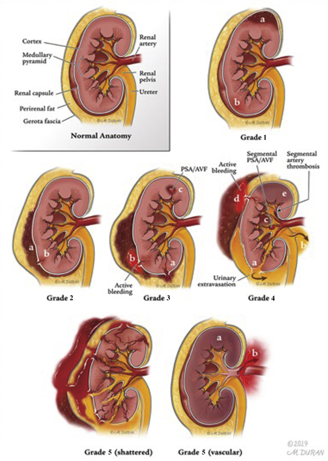
- Trauma: hemorrhage → distension of the capsule
- kidney infarction
Non-urologic:
- Muscle pain / Vertebral hernia
- Ac. appendicitis
- Spinal infections/injuries
- Cholecystitis
- Pneumonia (possible back pain)
- ectopic Pregnancy
- Endometriosis
- ovarian cyst torsion
- aortic aneurysm
- ileus
Renal colic
Pyelonephritis
Gerneral: Distended capsule/Distension at the level of the kidney
- kidney function:
- Crea (→Clearance)
- Urea, BUN
- Na + K (high K→arrhythmias)
- inflammatory markers
- Leukos, neutrophils,
- CRP
- ESR
- Plasma viscosity
- Gluc, Lac
- procalcitonin
- Coagulation, Thrombos
- INR
- Quick
- PT
- Anemia-marker, Thrombos, Hb, RBC, Ht
Caused by the inflammatory response (sepsis)
e.g. by UTI/Pyelonephritis
- Urine analysis: Proteins, RBC, WBC, Nitrate
- → then Urine cultures → follow-up antibiogram
yes, same as RBC at site of inflammation
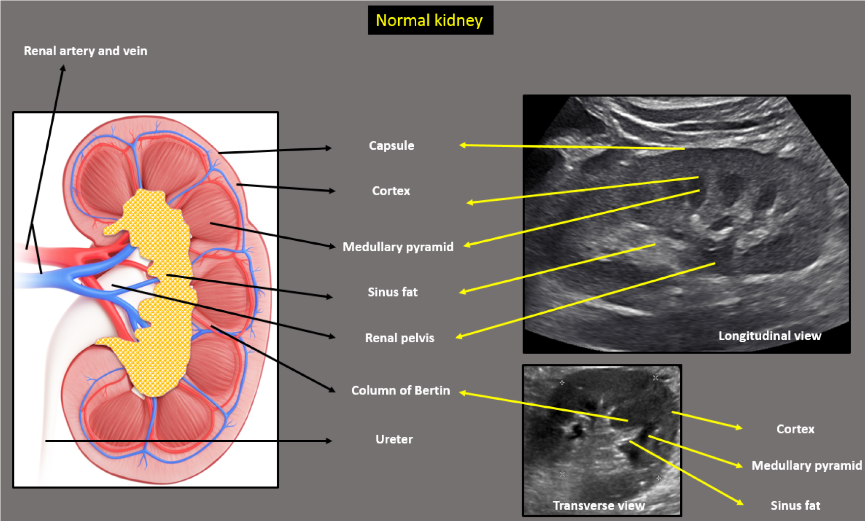
peripheric part hypoechoic: Cortex
→darker parts in the cortex=pyramids/medulla
central part hyperechoic: Pelvis + Calyx
- inc. adhesion platelest to vascular endothelium
- long term → vessel fibrosis
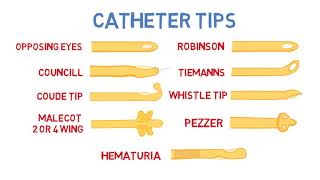
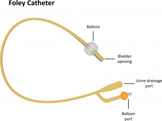
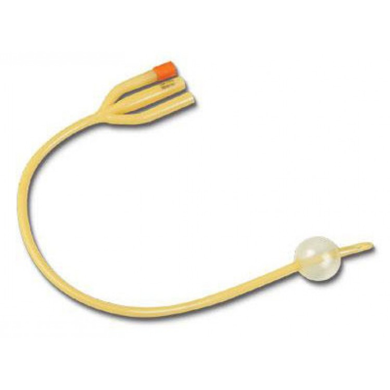
autostatic stays inside (balloon) = Foley CATHETER
non-autostatic just once = Malecot catheter
to flush with NaCl after surgery (anticoagulant)
silicon stays open
the rubber closes
when blood clots go through
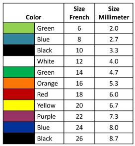
20ch,22ch
- bladder clot removal with saline
- blader evacuation
- instillation (chemotherpy?)
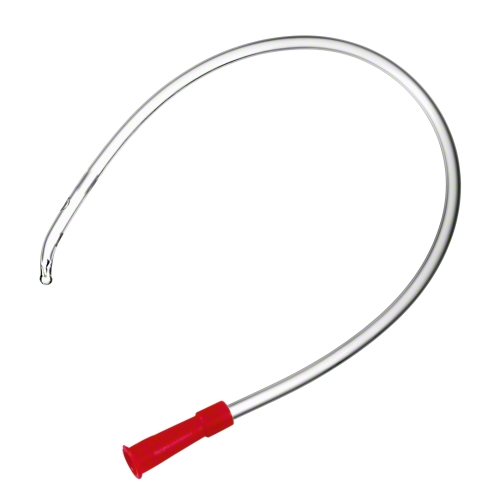
curved tip
- urethral stricture → move across it
- same es nelaton
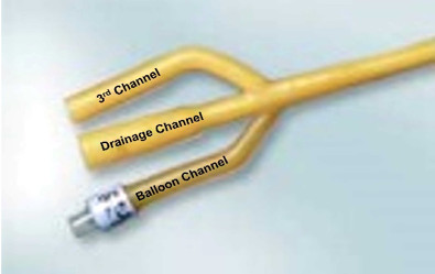
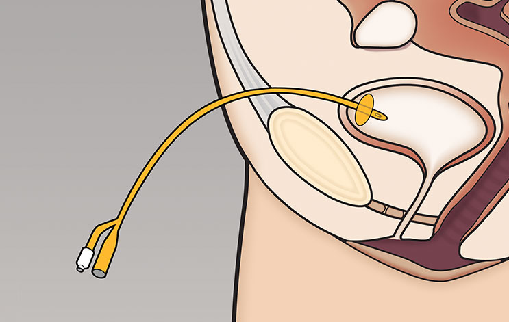
the balloon → fixes it in the bladder → autostatic
= only one which stays in the bladder
no color: urinary bag
colored part: indicated the size → saline can put into baloon (15-20ml)
- acute urinary retention
- chronic catheterization
- surgery
- endoscopic urologic surgery: Uretheral stent insertion, Uretroscopy[rigid/flexible] → no iregation !
no irregation if there is a suspected hydroneprhosis, obstruction,etc
→ can produce pyelonephritis due to more fluid insertion
Hematuria + Need for continous catheter
Post-endoscopic urologic procedures
→ TURBT + TURP (procedure → look it up)

ONLY WHEN BLADDER IS FULLY DISTENTED
- Trauma
- if you can't insert a intraurethral
Trocar 📷 → suprapubic penetration
2cm above pubic bone, perpendicular because bladder is distented und not behind the pubic bone
→ 📷
local anesthesia
check with a normal siringe + needle
Abrupt, firm "stabbing" motion
(slow constant pressure may push the bladder away)
Obstruction of the ureter
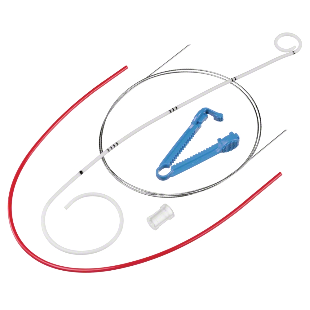
stent + guide wire
→ 📷
cystostocope with guidewirde into bladder → guide wire up intil the renal pelivis → insert the stent over the guide wire
double J → like a hook
🚑 Urologic emergencies
most common urologic emergencies
Cc: prostate massage 📷

- incr PSA
- inflammation: urine + culture
- Retention
- imaging: abcess, calcifcation, dilation seminal vesicals
Cephalosp
FluroQ
- discharge
- dysuria/urethral pruritus
- Clamydia
- Gonorrhea
- Trichomonas vaginalis
Doxy, Cephalo; Ceftriaxone + Azithromycin
⇒ SCARRING
congenital, inflammation, tumor, trauma (iatrogenic - TURBT+TURP)
- Inflammation
- ischemia
- trauma
- congenital
- malignancies
SCARRING due to inflammation/trauma/ischemia ⇒ decrease urethral diameter → obstruction
ant → corpus spongiosum lesion
post → urothelium → bladder neck
in conginital - no fusion of ant+post urethra
surgery(graft, open, etc), catheter, stent
- Enlarged Prostate BPH +ProstateCa
- Bladder cancer
- Urolithiasis
- Neurogenic cause (detrusor m., spinal cord injury)
- L-UTI-Trauma
most common cause for AUR in female
in women - pelvic floor collapses 📷 after gynecological cancer treatment, childbirth or heavy lifting.
In men- when prostate gland is removed.
- Sympatomimetic + anticholinergic drugs
phimosis - inability to retract forsjub
paramiphosis - inability to reduce forskin back → venous edema → "cock-ring-gangrene" / penile gland necrosis 📷
- Genitourinary procedures can result in failure to properly retract the foreskin after cystoscopy or bladder catheterization
- Penile trauma (piercing of genitals)
- infection
- Urinary retention
- Infertility
- Phimosis → Paramiphosis
- Condylomas
- Benign T + Maligncancies
→ Circumcision indicated
- retention or incontinence
- pain (abdominal, suprapubic, renal colic)
- less pronounced symptoms when chronic urinary retention (adaptive changes)

- pos. history for etiology?
- physical examination:
- palpation lower abdomen
- bladder palpation
- deep suprapubic palpation → discomfort
US of full bladder → let patient void → check bladder again
Volume ≥ 300cc → urinary retention
catheter urethral or suprapubic
2 way catheter
14-18 foley (french)catheter
- recent urological surgery
- trauma
- bacterial prostatitis
contraindic or failure to insert
+no manipulation and consequences of ureters + sphincter
-bowel perforation, wound infection
- Some benefits over indwelling urethral catheters:
- Prevent bladder neck and urethral dilatation, preventing urinary incontinence due to sphincter dysfunction.
- Assessment of the patient's ability to void before removing the catheter.
- Fewer infections.
- Avoid the risk of subsequent urethral stricture, a common complication in men requiring long-term urethral catheterization.
- Increased risk for complications: bowel perforation, wound infection.
due to rapid complete bladder decompression→
- hematuria
- increases fluid loss
- transient hypotension
- Postobstructive diuresis


etiology
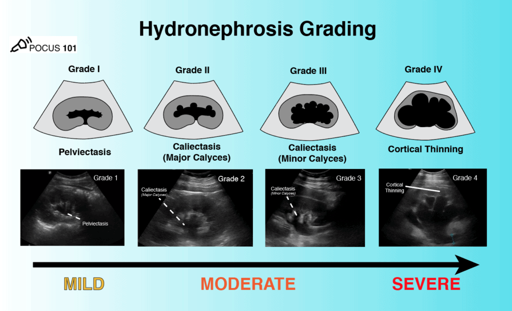
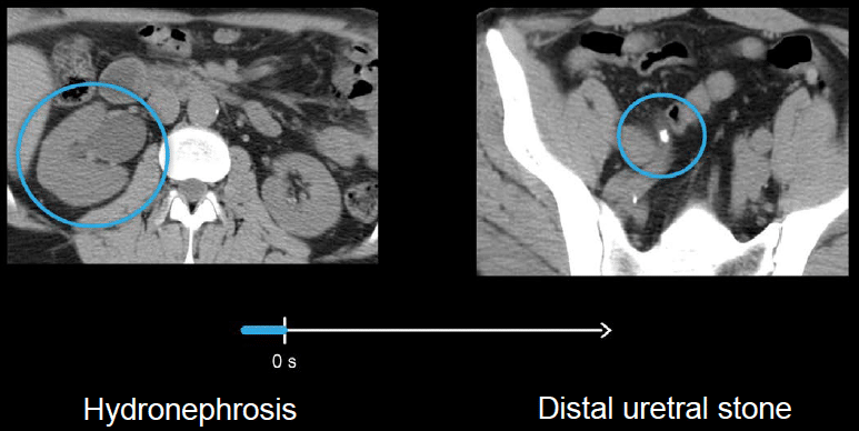
calyx/pelvis = hyperechogenic part → is pushed towards the cortex by urine = Hydronephrosis
- Stone in ureter (unilateral)
- stone in urethra if bilateral involvement
- tumors: in bladder, ureter or external compression
- Antibiotics → 3rd G. Cephalo., Fluroquinolnes
- Desobstruction + decompression
- Analgesic treatment:
- NSAID (!!!Nephrotoxicity)
- Acetominophen + Codein
- Opiods
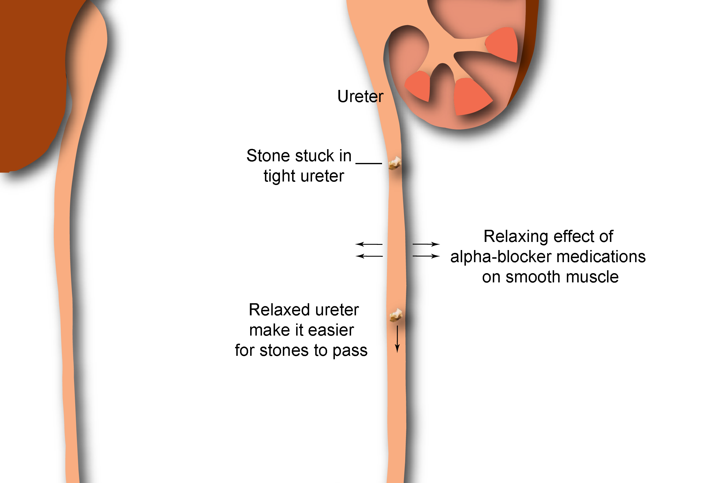
→ ESWL,MET (alpha- + Ca-Channel-Blocker),
→ Uretral stent, nephrostomy,
febrile renal colic → SEPSIS with huge inflammatory syndrome probably)
Admission Criteria
- Complicated renal colic
- Febrile renal colic (<36.5°C or >38°C, marked biological inflammatory syndrome)
- Hyperalgic renal colic (despite an effective dose of morphine)
- Rupture of the excretory path
- Acute obstructive renal failure (treat dyskalemia first)
- Pregnant
- stone in lumbar area of the ureter
- stone size >10mm
- complications (see above)
- emergency desobstruction:
- Urethral stent
- Nephrostomy
- elective stone destruction (after 3-4weeks)
Double J Stent Placement: https://www.youtube.com/watch?v=Wa20Csk7wJ8 Nephrostomy tube placement: https://www.youtube.com/watch?v=0PFKjNBt9xM
- Lower urinary tract obstruction (see above), Kidney stones
- UPJ obstruction/stenosis
- Vesicoureteric reflux
- pain - pos. giordano
- anuria(bilateral) - oliguria(unilateral)
- RF, HT, Hematuria
yes, 98% NPV
yes CT, false positive -26%
- dilation pc-system + ureter
- anechoic
- reduced parenchyma
- stones: hyperechoic + post-shadow
- suggestive obstructive cause by US → CT
- If nothing suggestive on US → go home
- if you cant visualize kidney on US → CT
- kidney marker Crea + BUN ; Ions → Esp K+
- CBC
- Inflammatory: Urine analyis → hematuria + pyuria; culture → possible UTI
BILATERAL
- only if obstruction bilateral
- or already kidney diseases present + unilateral obstruct.
⇒ will lead to crea increase
quick intervenstion!
- Tumor: Renal, Bladder, Prostate or external invasion
- BPH
- stones
- Infection: UTI nephritis
- trauma
- anticoagulation
dipstick + bga
hematuria?, infection?, anemia?, proteinuria?
- CBC + inflammatory markers (CRP, Gluc,
- BUN+Crea → RF? kidney marker
- Coagulation (INR, etc)
- Urine analysis + culture → nephritic, nephrotic, infection, RBC morphology, etc)
- tumor markers: PSA
- US,
- CT → abdomen+pelvis with contrast
- Cystoscopy
- also DRE → finger in po México

- Allergy to iodine
- chronic kidney disease: Creatinie → GFR<45!
- thyroid disease
- pregnancy
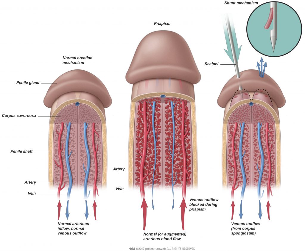
hard (>4h) but not horny
True disorder of penile erection:
- persists >4 hours
- beyond or unrelated to sexual interest or stimulation.
→ May occur at all age groups

impaired venous blood flow → venous congestion → low arterial inflow
95% of cases of priapism are due to ischemic cause
- pain
- absent or reduc. intracavernous arterial blood flow on doppler (no arterial flow)
- compartment sy (after 4)
- interstitial edema, sinusoidal endothelium destruction (after 12h)
- thrombocyte adherence due to endothelial dmg (after 24h)
- thrombosis, smooth muscle necrosis, fibroblastic transformation (after 48h)
- Neoplasm, infection, amyloidosis → vascular damage
- Neurogenic disorders, Vasactive agents → vasoconstriction
- hematological disorder (sickle cell, leukemia, multiple myelome etc) → eher stuttering priapism
corpora → hart
glans penis → soft
- Doppler (first)
- aspirate blood from corpora cavernosa → BGA
Source | pO2 (mmHg) | pCO2 (mmHg) | pH |
Normal arterial blood (room air) [similar values are found in arterial priapism] | >90 | <40 | 7.40 |
Normal mixed venous blood (room air) | 40 | 50 | 7.32 |
Ischaemic priapism (first corporal aspirate) | <30 | >60 | <7.25 |
trauma(perineal+ penil) → high flow fistula betw. artery + sinusoidal space
- Arterial is not fully rigid
- no pain
- sign of trauma
- Doppler → blood flow present turbular
- Blood aspiration → arterial blood
- consider CEUS
man with sickle cell
similar to ischemic
like ischemic
recurrency
- like ischemic
- alpha-adrenergic agonist
- erection inhibitors
The management of each acute episode is similar to that for ischemic priapism: aspiration/irrigation in combination with intracavernous injections of alpha-adrenergic agonists. The primary goal in the management of patients with stuttering priapism is the prevention of future episodes, which can be achieved pharmacologically.
gnRH antagonist
anti-androgen
phosphodiesterse Type5 inhibitors - PD5I
When the erect penis is forcibly bent, leading to a rupture of the tunica albuginea of the corporal bodies of the penis. Most common causes: sexual intercourse, forced flexion, masturbation, and rolling over.
- tunic rupture "Pop"
- immediate pain
- sudden loss of erection
- ecchymosis of the shaft + penile swelling = Eggplant deformity

- Paraphimosis
Necrotizin fascitis → of external genitalia + perineum
- Pain + swelling scrotum or perineum
- sepsis
- crepitus on palpation + foul smelling (if advanced)
- culture → whats the pathogen?
- CT or MRI → how is the extension? pararectal involvement?
- surgical debridement → should be as early + complete as possible otherwise → higher mortality + see picture below
- immediate empiric i.v. broad spectrum antibiotic
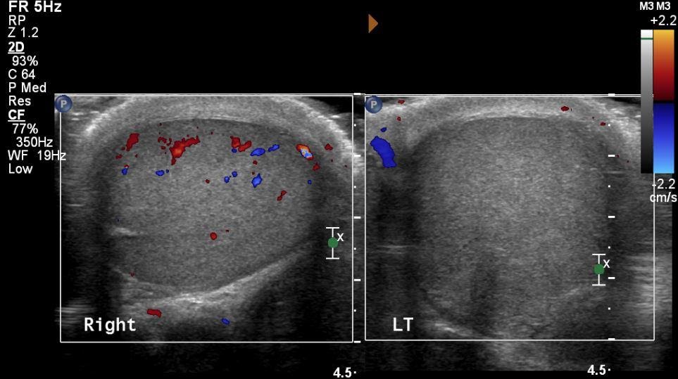
inadequate fixation of testis to tunica vaginalis 📷
CLASSIC FINDINGS: high testi, transverse long axis (instead of longitudinal)
- acute onset of pain (moderate-severe) → relieved by detorsion
- tenderness + swelling
- negative cremasteric reflex
- nausea, vomiting
- tendernes "knot" sup to testis
- Inflammation (epididymitis, orchitis,etc)
- Hydrocele
- Tumor
- Fournier's gangrene
- Trauma → rupture, hematoma
- Apendix torsion
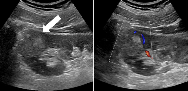
Doppler - no flow 📷
Successful detorsion is suggested by:
- Relief of pain
- Conversion of the transverse lie of the testis to a longitudinal orientation
- Lower position of the testis in the scrotum
- Return of normal arterial pulsations on Color Doppler ultrasound
>8h 📷
surgery
post. ribs
lumbar vertebra
damage to renal vascular
- Gray Turner's sign
- inspection → echhymosis, hematoma?
- palpation → might be tender, painfull
- dull percussion
- macroscopic hematuria
- Tissue damage?How about the function of the affected kidney?
- Fluid + Foreign bodies?
- Functional contralateral kidney?
- MRI → soft-tissue
- contrast CT → assessment of vasculature
- but US for free fluid
cystogram → hematuria bladder origin?
Retrograde urethrogram → urethral injury?
IVP
Angigraphy
not really used
Emobilization, staging
- myoglobinuria?? → might cuase ATN
- Normal BGA + CBC
- dipstick + analysis

- Gray turner (flank ecchymosis)
- tenderness - flank, abdomen
- hematuria (gross/micro)
- shock
after abdominal trauma → indicates renal trauma
even in minor renal trauma
absence does not exclude renal injury
Gross hematuria usually diminishes dramatically 2-6 hours after injury
- Sepsis
- Hemodynamic instability: Shock, Renal HT
- Expanding compression → rhabdomyolyiss + myoglobinuria
- renal atriopy, renal failure
trauma - iatrogenic
- often no symtoms - not in transection of ureter
- pain only when obstructed
- RF
- hematuria
- surgery
- AB
- ureterostomy 📷
- irrigation + drainage → in retention
- Stenting → in retention
- fistula
- ureteral stricture →Hydronephrosis
- infection
- Infection - fever, leucos, sepsis, wound infection
- flank mass
- lleus, urinary fistula (to skin/vagina)
- urinoma (encapsulated fluid collection due to urine leakage)
full bladder → geh pissen bevor du auto fährst
- free fluid on US
- abdominal trauma → suprapubic pain
- pelvic fracture
- cant void, despite the urge
- gross hematura
- Rebound tenderness
- Abdominal guarding, rigidity
- displacement of prostate
Extraperitoneal | Intraperitoneal |
Urine found in umbilicus,
anterior thighs, perineum
• Dysuria
• Hematuria
• Suprapubic swelling, redness.
tenderness | Occurs with penetrating or
blunt rupture of distended
bladder
• 15-45% of bladder trauma
• Urgency and inability to void
• Signs and symptoms of
shock
• Abdominal distension |
⇒ diagnosed with cystogram
- high mortality association!
- death due to hemorrhage, sepsis or anrectal injury
- Bladder drainage
- cystorrhaphy (suture bladder)
open → get the blood out → bladder sutures
- IVP
- Urethrogram
- CT
→ double dose exr. urography
- pelvic fracture in posterior associated
- ant - isolated injury
Sudden deceleration injuries (bladder shears off urethra)
- pain
- unable to urinate
- hematuria
- perineal bruising "butterfly pattern" 📷
- displaced prostate
- Scrotal hematoma
- rebound tenderness + abdominal rigidity
- impotence+ incontinence,sepsis - post
- stricture - ant
Posterior:
- Permanent impotence
- Permanent incontinence
- Cellulitis
- Sepsis
- Urethral stricture
Anterior:
- Urethral reconstruction can have reanastomosis defects
- Urethral strictures
- Infection from extravasated blood or urine, which can lead to necrosis
urethrovaginal communication
🪨 Urolithiasis
fused version with renal lithiasis in Nephrology
crystalline mineral deposits → migrate + obstruct ureter → renal colic pain

Classic: 💥Colic wave pain+ 🩸Hematuria
severe: UREMIC SYNDROME + SEPSIS
can vary widly:
- inc. urgency + freq
- diffuse abdominal pain
- nausea
- testicular pain
- uremic syndrome - in uric acid stone
= site of pain
- Diet
- reduced water intake
- disorders leading to high Ca
- HT
- DM
- calcium oxalate - most common 75%
- calcium phosphate - 15%
- uric acid - 10%
- struvite, cysteine - rare
- hypercalciuria + oxalaluria due to any cause (hPTH)
- obstruction of the ureter or UPJ
- animal protein intake, vitC + D intake
🟡 Urease-producing germs = Proteus + Klebsiella → only in upper UTI
- Ions (Ca, P)
- Urea + Crea
- Uric acid
- PTH
- CRP, WBC
- Hb, Hct
- Dipstick - 🩸 Hematuria
- 🧫 Infection (Leu, nitrate,ph,culture)
- Pain control → NSAID, Opiates, warming,
- MET: alpha-blocker + ca-channel blocker (see below) 📷
- UTI + Sepsis → AB
- Prevention + Recurrance
- Diet →VitC, animal prot, Na, Ca, purine in uric acid
- sufficient Fluid intake → 2.5-3L/day
- sodium restriction if high excretion
- Drugs → thiazide
- Lifestyle advide → normal BMI
- treat Hyperparathyroidsm
- Urologic intervention
signs of sepsis!! → Urosepsis
no delay it until sepsis is resolved
→ exception: sepsis + abscess
- urine culture
- urine microscopy
→ exclude UTI / treat prior to removal therapy
→ give AB prophylaxis
evtl. stop antithrombotic therapy
Infections, refractory pain, ↓ renal function
- Shock Wave Lithotripsy
- offer stenting / perc. nephrostomy
→ see below
👉🏽 📹
renal + prox ureteral stones
stone density >1000HU
stone >10mm
- pregnancy
- bleeding diatheses
- UTI
- anatomical obstruction distal to stone (e.g. ureteral stricture)
increase power session by session
ureteral + renal stones >10mm
👉🏽 📹
ureteral trauma + residual fragments
uncomplicated cases, no trauma, stone-free procedure
renal stone >20mm → percutaneous nephrolithotomy
👉🏽 📹
→ Precise pssessment of the stone with contrast (redrograde/CT)
calyx tear
- Ultima ratio - in rare cases, when others fails
- UPJ obstruction → concomitant reconstructive surgery
uric acid stones → alkalinization of the urine with Sodiumbicarb. i.v.
🧫 UTI
fused with UTI - Nephrology
- outside hospital aquired + hospital aquired
- sensible germs + resistance germs
bacteriuria + leucocyturo
→ repeat exam!
could be do to contamination of sample or contamination via catheter
- other inflammatory causes → stone, tumor, irradiation damage, nephrologic D
- atypical germs

G- → E.coli, Klebsiella, Enterobacter, Proteus
G+ →Enterococc(Klebs, Pseudomonas), StaphAureus
*most common - assiciated with infection outside hospital, others are nosocomial
- Neisseria gonorrhea
- clamydia
- staph strep
cloudy 📷, bad smell, +/- hematuria
dipstick (leukoC-esterase, nitrite)
- Disruption urinary flow due to obstruction
- neurologic bladder + sphincter abnormality
- vesicoureteral reflux
- foreign body (catheter, ureteral stent, nephrostomy)
- postmenopausal
- pregnancy
→ When SEPSIS → check for obstruction/Abcess → treat
CT: cant detect: gas-forming abcess, hemorrhage, obstruction, anatomic factors
- pregnancy
- pre-urologic procedure
- reflux
- obstruction
- neutropenia
- recent transplant
acute pyelonephritis in 25-40%!
→premature delivery, low birth weigh, baby mortality
! SCREEEEEEENING bidde
Amoxi-Clavu or Phosphomycin
no Fluros, no TMP in first trimester
all have bacteriuria → absent/minimal symptoms
Remove catheter, do intermittent cathetering, only treat when symptomatic (fever,dysuria)
malformation
- intercourse
- bowel disorders
- contraceptives
- gyn-infections
- foreign body in urethra+bladder
e.coli
pollakiuria (freq. mictuition of small amount)
dysuria - burning
⇒ lower urinary tract signs
cloudy urine
+/- hematuria
fever, lumbar pain → acute pyelonephritis
dipstick - leukos+Nit
culture
- AB
- analgesic - NSAID or more
FluroQui
- TMP-SMX
- Norfloxaxin
- Amoxi + clavulanic acid
- FluroQui
- onset of symptoms
Acute:
- systemic inflammatory response → fever, Np
- AUR
- Urinanalysis Nit +
Chronic
- no acute phase reaction
- local symptoms
- Leu in semen+germs
abacterial chronic prostatitis
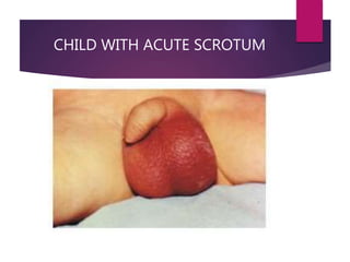
Testicular Ca + Torsion
like APN but with acute scrotum 📷
secondary to acute or chronic bacterial prostatitis
- sterile leucocyturia
- cloud urine, chronic
- extreme pollakiuria
- small bladder
- Forunier gangrene
bacterial inflammation of PC-epithelium + renal parenchyma
- obstructive
- non-obstrive (reflux, attypical)
obstruction → impaired urine flow → ascending bacterial infection + microabcess
- colic pain
- sign of systemic infect → Sepsis (HypoT, TC, TPn), fever, chills
- lower urinary tract sign (dysuria, pollakiuria, freq.)
- lumbar flank pain, costovertebral tenderness - giordano
- pyuria
- history of recent cystitis
- nausea + vomiting
qSOFA ≥2 suggests a poorer outcome & alert for possible infection (when previously unknown)
- CBC
- Crea + BUN, Electrolytes
- BGA
- inflammatory markers: Lac, CRP, Glu
- Bili + Coagulation test
- Urinanalyis + culture (2 sets)
- IV AB
- FluroQ
- Aminoglycoside (+ amoxi)
- Cephalos
- Pathogen specific - Vanco, linezolid
- Give Volume
- Address Electrolyte abnormalities
- Decompression of obstruction (see Hydronephrosis)
- cant get through in stable pat
- unstable patient
- pyonephrosis (pus)
DM, Immunosupressed, eldery
DM, posttransplant kindey
- Atropy + scars
- HT + CKD
- Renal abcess
- papillary necrosis
- perinephric phlegon
- pyonephrosis
- septic shock
hematogenous , staph
high fever, back pain, nephromegaly
urineanalyis
US
CT
perinephric phlegmon
no improvement on AB therapüy
pus outside the renal capsule
back pain US + CT
purulent destruction renal parenchyma → nephrectomy + AB needed
- Procalcitonin → incr. in big amount of bacterial, fungal + parasitis
- AB asap!
→ start empiricial broad spectrum early → then adapt to antibiogram
- give volume
- vasopressor (NE,dobutamin)
- evtl. cortisone in renal failure
🦀 Onco-Urology
General + BPH
- Urinananalysis
- Urine culture
- Urine sediment microscopy
CBC
Urea, Crea, K, Na
Gluc
Coagulation
- kidney
- prostate
- bladder
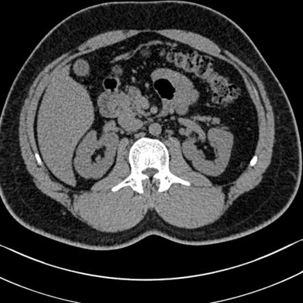
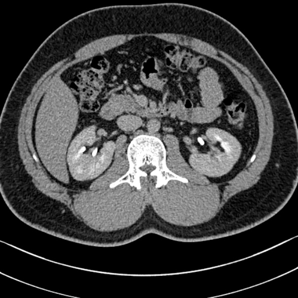
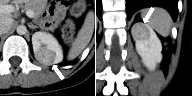
mass in the inf. pole of the kidney
→Tumor?? → do CT
- pregnancy
- CDK: GFR < 45
- allergy
- ...others
Urologic:
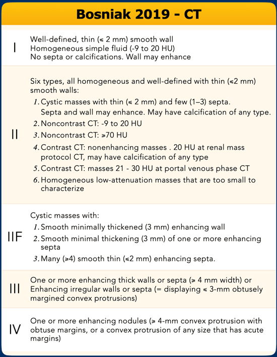
- parenchyma mass: cyst (bosniak classif.) or solid tumors
- tumor:
- benign(oncocytoma or AML most common) or
- malignant (carcinoma with renal cells → most common?
- calyx/pelvis mass: urothelioma
- abcess → CT: inhomogenous kidney / patchy hypointense areas → give AB + drainage
- subcapsular hematoma
- hydronephrosis / UPJ-synd.
- Polycystic kidney diseases
Non-Urologic:
- aortic aneurysm with rupture into retroperito.
- metastasis
- adrenal tumor
- sarcoma
- adenopathies
nephrectomy
Neproureterectomy
- biopsy,
- surgery nephrectomy→ partial or radical
- surveillance
- ablative techniques
- stage of the tumor → dimension
- fitness → in young:
- oncological: nephrectomy
- functional:
Risk score during surgery:
Post-OP-Complication + renal sparing potential
- polar location? → 1 point
- exophytic rate: mainly outside → 1 point
- Rim location: at the medial side of the rim → 2 points
- Renal sinus involvement: near it → 2 points
- Urinary collective system involvement: invades calyx → 2 points
- Tumor size: <4cm 1point; 4-7 2 points; >7 3 points → 2 point
⇒ 10 points
⇒ 6-7 partial nephrectomy
invasion of the pelvis
laproscopic
robotic
open
- anamnesis → medication
- allergies
- complete imaginistic
- complete biological evaluation: CBC, renal function, coagulation
- Vital paramenters
- Checkup! Is it the right person?? Ask! → presurgical checkup
wound care → infection!
pain management (morphins, Nsaid, acetominophen)
drain tube
vital parameters
thromboprophylaxis
mobilisation
ERAS
LUTS
- symptoms of LUTS - dysuriua, weak stream, hesitating, terminal dribbling
- Polyuria
- bladder overactity → hyperexitability, fibrosis, contractibility ⇒ urgency urinary incontinence
- US → Postmicturial residue
- Uroflowmetry → specific pattern
DRE 👉🏽
- Infection: Urinalyis, culture, Glu, blood
- renal marker Crea + Urea
- PSA
Prostate out, symptomatic, Drugs
- symptomatic
- subvesical obstruction surgery → retropubic/transvesicular open surgery, endoscopic - transurethral resection 📷, laser
- lifestyle change
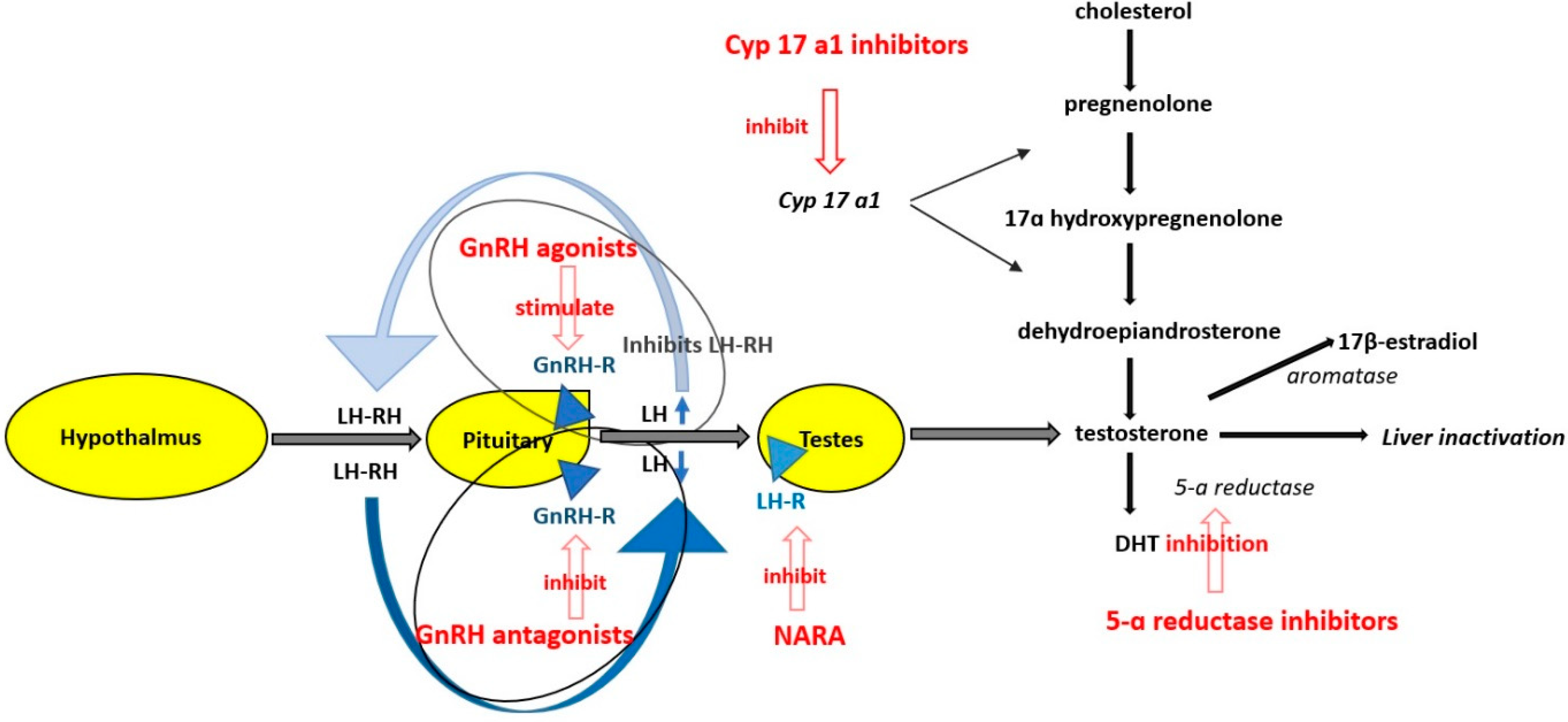
- drugs: 📷
- 5-alpha reductase inhibitor (5ARI)→ reduce androgens
- Alpha1-blocker + antimuscarinics →relax bladder+prostate
- PDE5 inhibitor → correct erectile dysfunction
- Desmopressin

5-alpha reductase inhibitors (5-ARIs) effectively reduce the serum and intraprostatic concentration of DHT, causing an involution of prostate tissue
A phosphodiesterase type 5 inhibitor (PDE5 inhibitor) is a drug used to block the degradative action of cMP-specific phosphodiesterase type 5 (PDE5) on cyclic GMP in the smooth muscle cells lining the blood vessels supplying various tissues. These drugs dilate the corpora cavernosa of the penis, facilitating erection with sexual stimulation, and are used in the treatment of erectile dysfunction (ED)
- bleeding
- TUR-syndrome → Fluid instillation during Transurethral resection (TUR) → hyponatriemic hypervolemia
- Bladder neck stenosis
- Urethral strictures
- Urinary incontinence
- UTI

Malignant lesions

PSA, DRE, US abdomen
- age (>50)
- genetic
- initial PSA → >1ng/ml at 40 years / >2 at 60 years [normal value: 0-4ng/ml]
- Genes: MLH1, MSH2, MSH6, PMS2 (Lynch Syndrome), BRCA1, BRCA2, ATM, PALB2, CHEK2.
- Genetic testing: Positive family history, high-risk prostate cancer, Ashkenazi Jewish ancestry, intraductal histology.
→ diagnose prostate cancer in early stage → treatment (prophylactic) with low side effects
SLOW! (microscopic → extracapsular → >5y: diagnosis)
find before 5 years! only with screening
after 5 years: - presentation in advanced stage
- Asymptomatic
- Signs of local extension:
- pain
- mictuition difficulty
- pollakiuria + dysuria
- hematuria
- hematospermia
B-symptomatic (if systemic)
weight loss asthenia anorexia sciatica pseudo-seizures signs of renal failure obstructive jaundice neurological changes
ivory verterbra back pain 📷
Parameter | Total PSA |
Sensitivity | 87% |
Specificity | 15% |
0-4 ng/ml
PSA inc. or abnormal DRE
- PSA>4,
- PSA-incr.with >0,75/year
- abnormal DRE
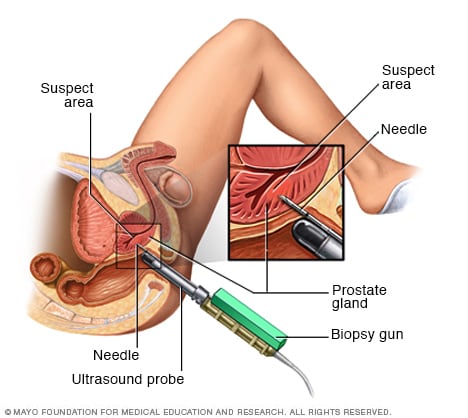
→ US-guided transrectal/perineal
- BPH (related to age)
- Prostatatis + UTI
- external manipulation of prostate - e.g. DRE (=DRU)
T1 - no change (not detectable with DRE)
T2- hard, not beyond capsule
T3- beyond capsule
T4-bulky prostato-pelvic tumor fixed to pelvic wall, infiltration rectum
if NV-bundle 📷 is damaged → loosing the potency of the patient if cancer has developed in the later part
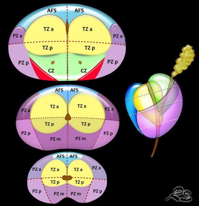
peripheral zone: 60-70%
Transitional zone: 20-30% (here does also BPH develop→uretral stenosis)
anterior-fibrular muscular stroma: 5%
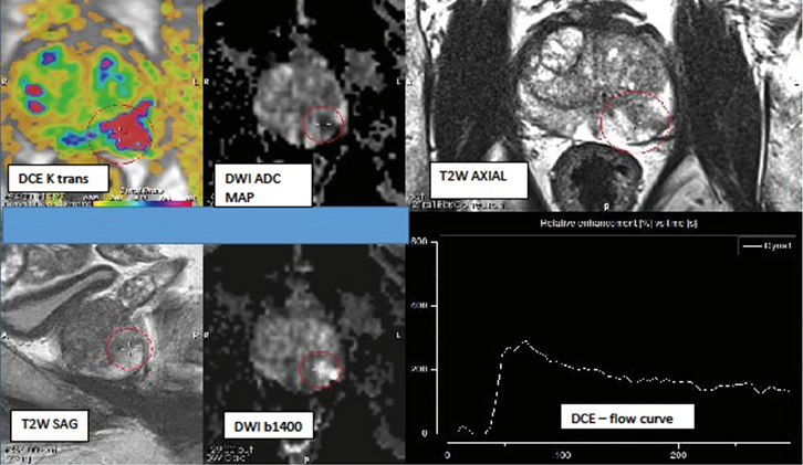
- T2 → look for hyposignal area in peripheric zone → than look for transitional zone
- Difusion weighted: Functional → cellularity → if hypercellularity → more water → hypersignal in DWI + hyposignal in adc
- ADC → functional → hyposignal in hypercellularity
- Contrast: early enhancement
AFMS-tumor?
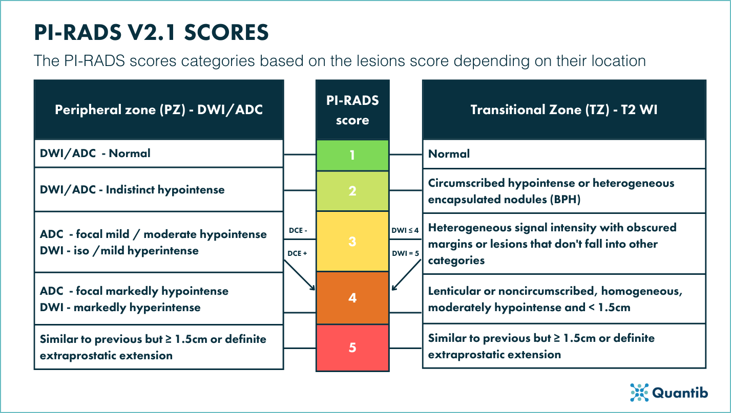
Clinically significant: 3-5
insignificant: 1-2
3: 25% Clinically significant PCa
4: 50-60%
5: 90%
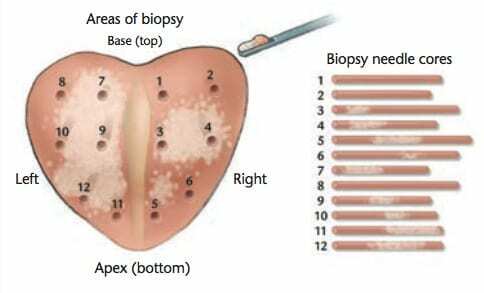
take 12 biopsy fragments out of every peripheric zone 📷
Malign/Benign
- Type of PCa
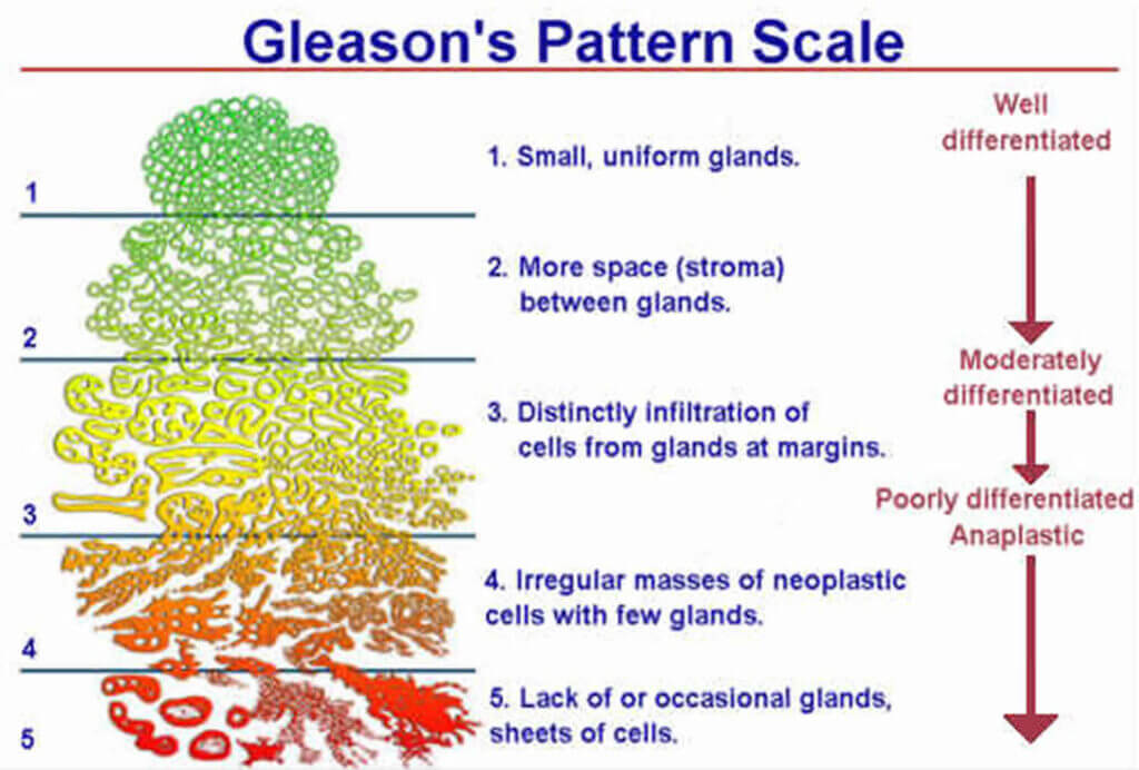
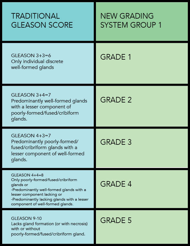
- Grading
- Number of positive cores
- Length of cancer
→ Criteria for insignificant PCa
- No Gleason patter of 4 or 5 / Gleason sum <7
- <3 pos. cores (12 samples from different area)
- <50% positive core involvement
⇒ otherwise = significant PCa
DRE, finger in po mexiko ⇒ DRE CLASSIFICATION
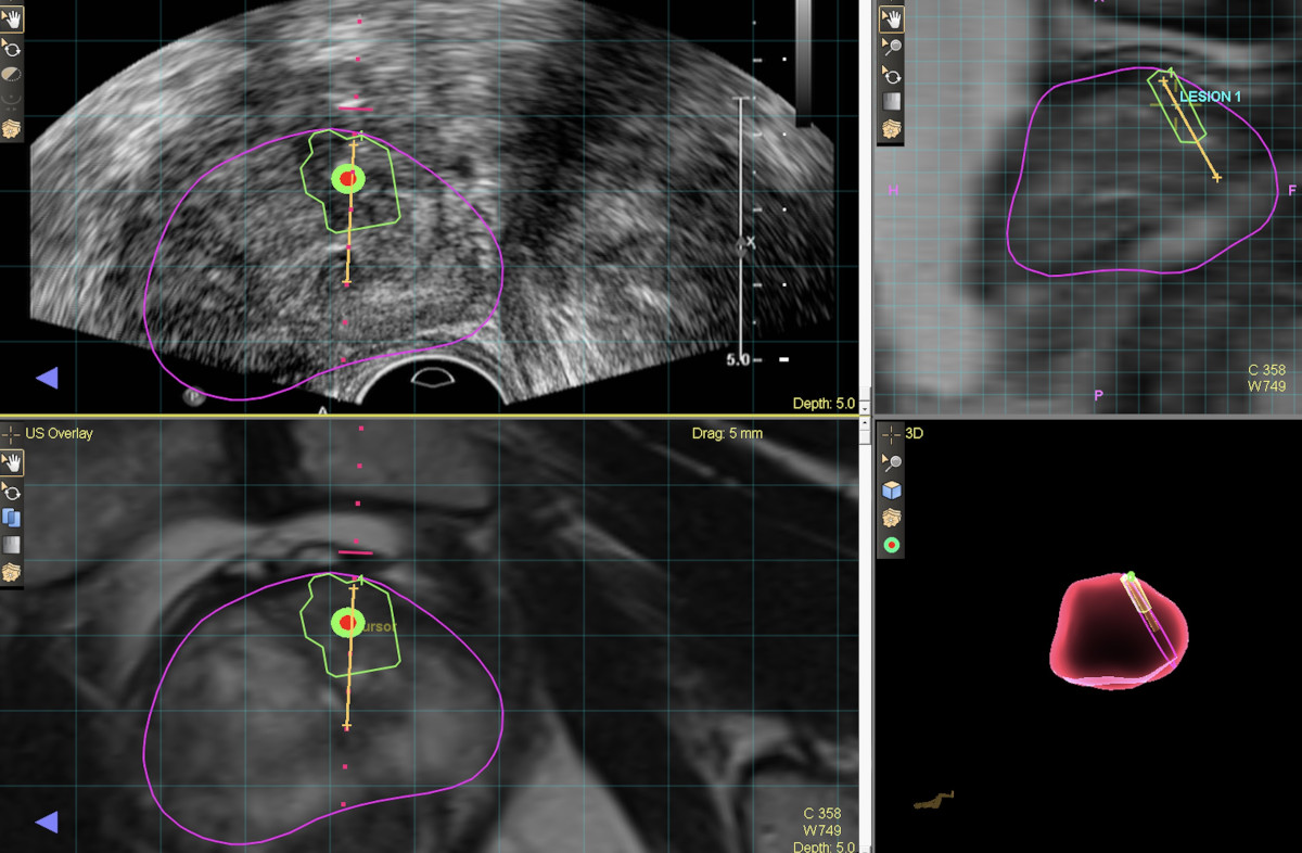
oder mpMRI
TRUS (transrectal) + elstography
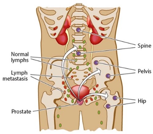
for regional
pelvic LN
mpMRI or CTpelvis + contrast
- NX: Regional lymph nodes cannot be assessed.
- NO: No regional lymph node metastasis.
- N1: Regional lymph node metastasis.
Scintigraphy (bones)
CT abdomen
- M0: No dist. mets
- M1: Distant mets
- M1a: Non-reginal LN mets
- M1b: Bones
- M1c: Other sites
PSA >10, bone pain, Gleason 8-10, T3-T4
biochemical recurrence = high PSA during follow up
PSA < 10,GS < 7, T1-T2a → low risk
PSA >10,GS >7,≥T2b → intermediate/high risk (high >20)
→ low risk
intermediate(=7)/high risk (>7)
preffered: active surveilance
alternatives:
- organ preserving
- focal therapy
- brachytherapy
- radical
- prostatectomy
- radiotherapy
- insignificant PCa: see Epstein criteria
- short life expectency (<10 years)
Epstein criteria:
→ Criteria for insignificant PCa
- No Gleason patter of 4 or 5 / Gleason sum <7
- <3 pos. cores (12 samples from different area)
- <50% positive core involvement
• PSA • DRE • mpMRI • Prostate biopsy
high intensitiy focused ultrasound → heat → tissue destruction
Radiotherapy ≤ T2N0M0, patient who dont want surgery, can be done directly after prostatectomy in T3
Hormonal: T3,T4
T1a + Life expactation <10years
HIFU, brachytherapy
Radio + radical prostatectomy
- External radiotherapy
- Hormonal treatment
- Excision of prostate
- Lymphadenectomy (lymphadenectomy not always needed! → only when chance of involvement is >5% (use calculator)
- Vesico-urethral anastomosis
- oncological → radical removal
- preserve continence → sphincter (90% normal after OP)
- preserve potency → NV-bundle (bilateral nerve sparing → 70% keep potency, unilateral nerve spacring → 40%, non-nerve sparing 10%)
- bleeding
- thrombosis + UTI, fistula
- late: impotence, incontinence, strictures
residual tumor
- erectile dysfunction
- incontinence
N+ M+
→ Antiandrogen
old
or young with HPV + other STDs
can be cured in 80% if no Metastasis
penis presevering surgical remove
- topical treatment + abrasion
- partial amputation, wide excision + reconstruction(glansectomy +resurfacing)
- chemo + radiotherapy
- lymphadenectopathy
- Adrenal tumor
- Urothelial tumor
- Urinary bladder tumor
⚡ Neuro-Urology
Neurogenic Lower urinary tract dysfunction
inter sphi. only sympath. control
SNS | PSNS | |
Detrusor | (β3 receptors) - | (muscarinic receptors) + |
Internal sphincter (smooth) | (α1 receptors) + | + |
freq, urgency, incontinence
→ hestance, weak stream, imcomplete voiding, dribbling
Stroke, Dementias, Parkinson, brain tumor, cerebral trauma, cerebral palsy
- urgency, intcontinence
- overflow incontinence
- no bladder sensition
spinal trauma, spina bifida
RETENTION without obstruction

- lumbar spine lesion, degenerative diseases, disk prolapse, lumbar canal stenosis
- Periph. neuropathy: DM, alcohol
- Iatrogenic pelvic lesions
bladder dysfunction → UTI + chronic RF
- posttrauma
- urinary retention → infection, hydronephrosis
- extremly enlarged bladder - high pressure
- reduced renal function

Sensation S2-S5 both sides) Presence (increased/normal/reduced/absent) Type (light touch/pin prick) Affected dermatomes Reflexes (increased/normal/reduced/absent) Bulbocavernous reflex Perianal/anal reflex Knee and ankle reflexes Plantar responses (Babinski) Anal sphincter tone Presence (increased/normal/reduced/absent) Voluntary contractions of anal sphincter and pelvic muscles (increased/normal/reduced/absent) Prostate palpation Descensus (prolapse) of pelvic organs
- US
- Uroflowmetry
- Cystomanometry → messure bladder P
- Cystouretrography
- Urine analysis
no
- continence
- protection of UUT-involvement
- restoration of LUT function
- decompression, complete +periodically → less UTI, kidney protection
- Drugs:
- Antimuscarinic, beta3 agonist → if patient has incontinence
- a1 blocker, botox → if patient has urinary retention
- stent, catheter
--- | SNS | PSNS |
Detrusor | (β3 receptors) - | (muscarinic receptors) + |
Internal sphincter (smooth) | (α1 receptors) + | + |