
Table of content
- Ophthalmology
- 👁️🗨️ TR QBank
- 👁️ Eye anatomy & Visual function
- 🪞 Refractive errors
- 👓 Disorders of binocular vision
- 🏐 Pathology of the orbit
- 🥠 Eyelid pathology
- ☄️ Conjunctiva pathology
- 💧 Lacrimal pathology
- 🦏 Cornea pathology
- 🌈 Uveal pathology
- 🥚 Lens pathology
- 🚧 Glaucoma
- 🕸️ Retinal pathology
- ⚡ Optic nerve pathology
- 🕳️ Pupil pathology
- 🔪 Ocular Trauma
Member Resources
 Year 4
 Year 5
 Year 6
Ophthalmology

👁️🗨️ TR QBank
History
History of present illness (HPI)
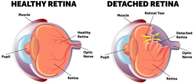
retinal detachment + tears 📷 📷
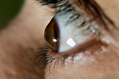
Floaters look like small specks, dots, circles, lines or cobwebs in your field of vision. While they seem to be in front of your eye, they are floating inside. Floaters are tiny clumps of gel or cells inside the vitreous that fills your eye. What you see are the shadows these clumps cast on your retina. Flashes can look like flashing lights or lightning streaks in your field of vision. Some people compare them to seeing “stars” after being hit on the head.
young: migraine vessel spasm
elderly: micro-emboli
📷 ⇒ ischemic event or retinal detachment
- always?
- worsen when reading/watching TV? → dry eyes, blink less
- glare problem at night? → cataract
- DM present? → poor control

- hows the pain? → scratchy, aching, only with bright light
- discharge? → infection
- allergies
- blepharitis (eyelid infection)
temporal (giant cell) arteritis
→ also jaw cladication, polymylagias, weight loss, night sweats
DD: migraine
"Right Hand Column"
- DM
- HT
- Coronary artery disease
- thyroid diseases
- asthma → in case of beta-blocker prescription, might trigger bronchospasm

FH (family history): glaucoma or blindness?
Allergies: esp. sulfadrugs (ingredient in a lot of pressure releasing drops)
Medication - eyedrops? anticoagulants? oral betablocker?
"Vital signs"
- Vision
- Pupil
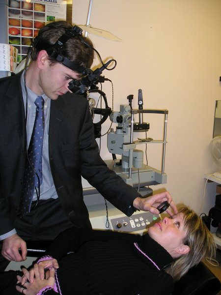
- Tonometry (Pressure)
BEFORE DILATING PATIENTS EYE! → the drops will mess up these parameters
Visual Acuity + Pressure
Far vision
Snellen chart 📷
refractive error
pinhole 📷
filter misaligned rays → central ray on retina
think about cataracts or other media opacities
- opacification - caterac, corneal edema, vitrous hemorrhage, etc)
- optical n. + retinal illness: retinal detachment, diabetic retinop, ischemia, toxic, ...
- Amblyopia → without organic cause (strabismus)
Near vision
near-card or small print in newspaper
>40y → Presbyopia (Alterssichtigkeit)
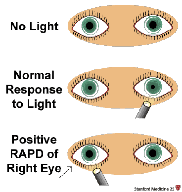
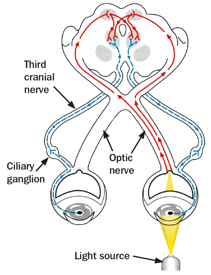
Pupils
constriction → also the non-illuminated one (equal and synchronous)
constriction
= Marcus Gunn Pupil → one eye cant sense light as good as the other one
⇒ Swinging light test 📷: abnormal: dilation of both eye when flashing in the direction of pathological eye and constriction when going back in the direction of the good eye
Pressure
how much force is necessary to flatten an predetermined area of corneal surface
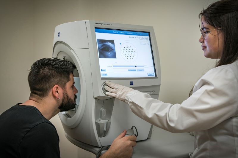
Confrontational fields
visual field (periph. vision)
"tunnel vision" due to neurological diseases or glaucoma
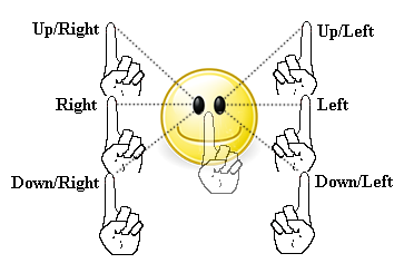
Extraocular movements (EOMs)
- you might see decr. mobility
- or you see nothing but pat. complains about double vision
mono - or biocular diplopia
→ cover one eye? → still double vision → monoocular → refractive error: astigmatism, cateract, corneal surface wrinkling
→ if double vision is solved after cover one eye → biocular diplopia → neuromuscular paralysis or posttraumatic muscle entrapment → determine what muscle group/ nerve is involved
(dont be confused it will be covered later again)
Slit Lamp exam
- External (EXT)
- Lids and lacrimation (L/L)
- Conjunctiva + Sclera (C / S)
- Cornea (K)
- Anterior Chamber (AC)
- Iris (I)
- Lens (L)
- Vitreous (V)
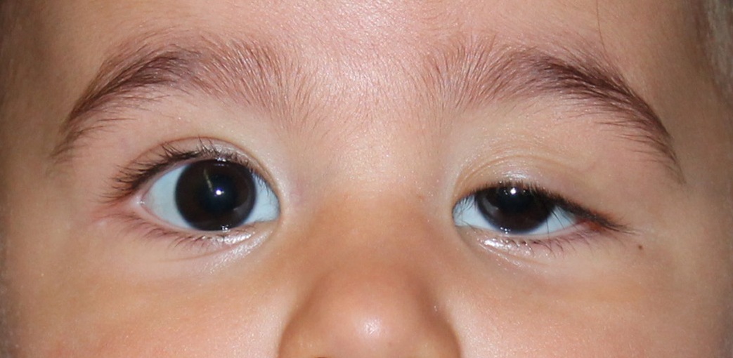
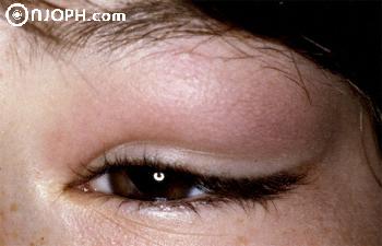
→ Ptosis (drooping up. eyelid 📷) or Proptosis(=Exopthalmus 📷)
→ Conjunctivits? → swelling pre-auricular nodes (in front of the ear) and sub-mandibular/mental nodes?
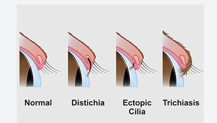
→ Blepharitis? (infection lid margins) 📷
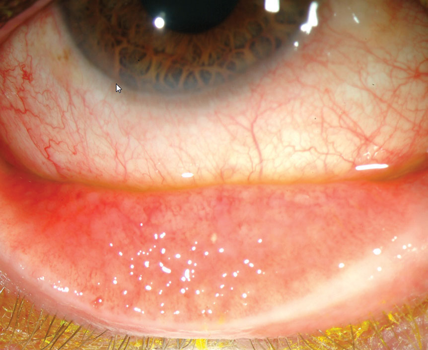
→ follicles or papillary bumps on the lid inside? 📷→ infection / irritation
→ white, icteric?
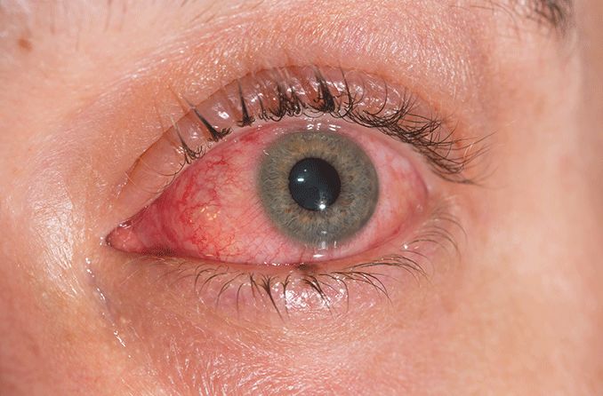
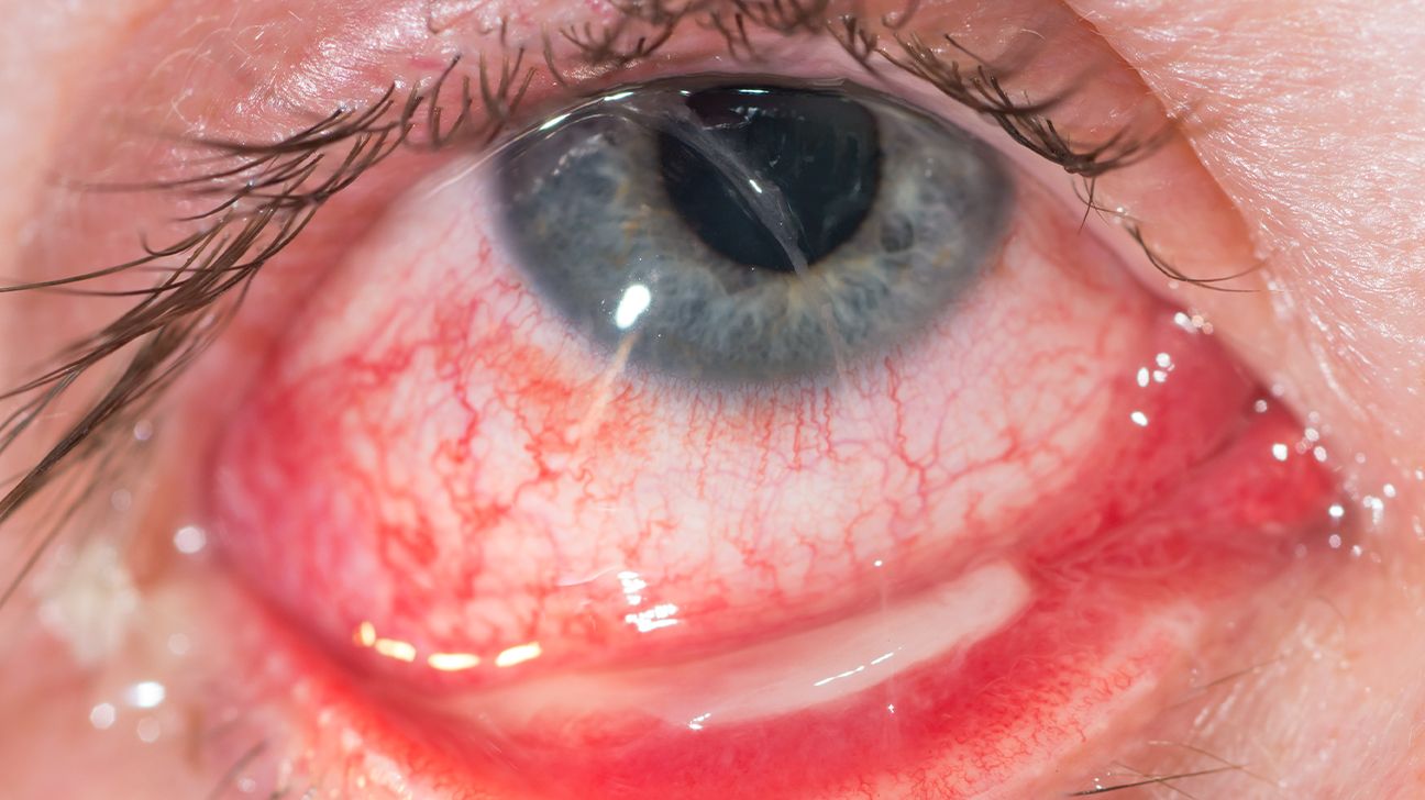
→ injected conjunctival blood vessel (red + inflammed) 📷
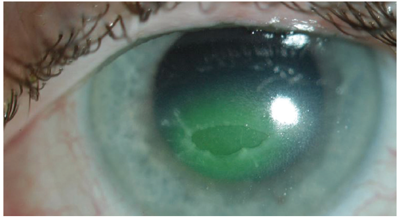
→ Corneal erosions + abrasion? 📷 → trauma → consider Fluorescein dye 📷
→ Clear stroma?
→ folds or gutatta bumps at endothelial surface? 📷
→ individual cells (PMN) or protein flare? 📷 📷→ inflammation (uveitis, iritis, postsurgery)

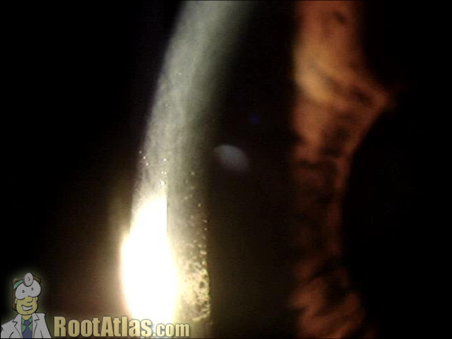
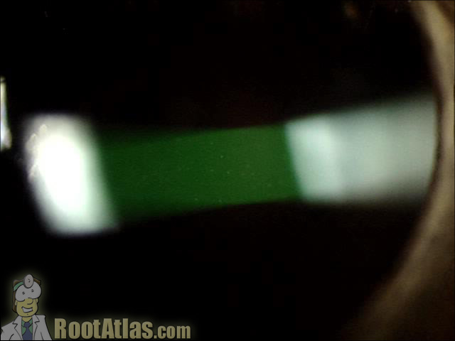
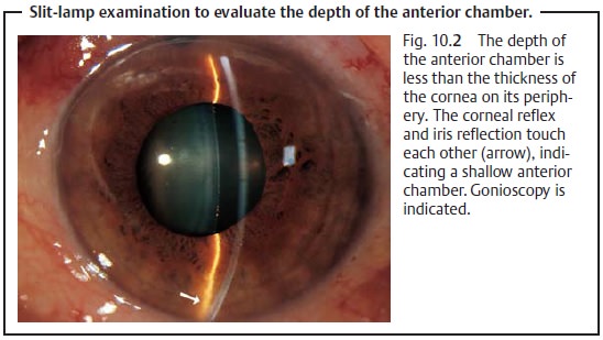
→ shallow ant. chamber 📷→ setup for angle-occlusion glaucoma!
→ flat? , pupil round?
→ if diabetes or old retinal vascular occlusion → neovascularization?
→ clear or hazy with cateract? 📷
→ phakic, pseuophakic, aphakic?

→ floating cells? → retinal hemorrhage or detachment
Dilated Fundus Exam
Basics
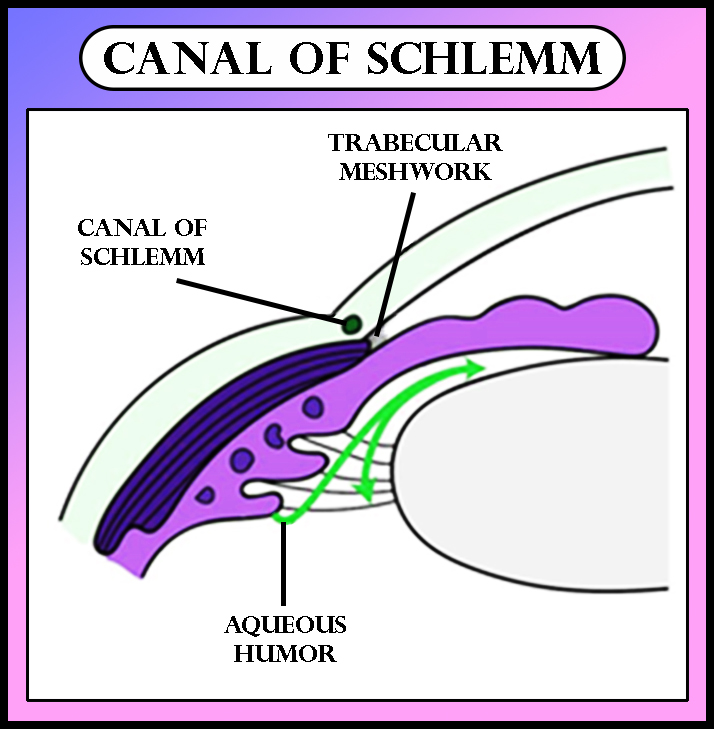
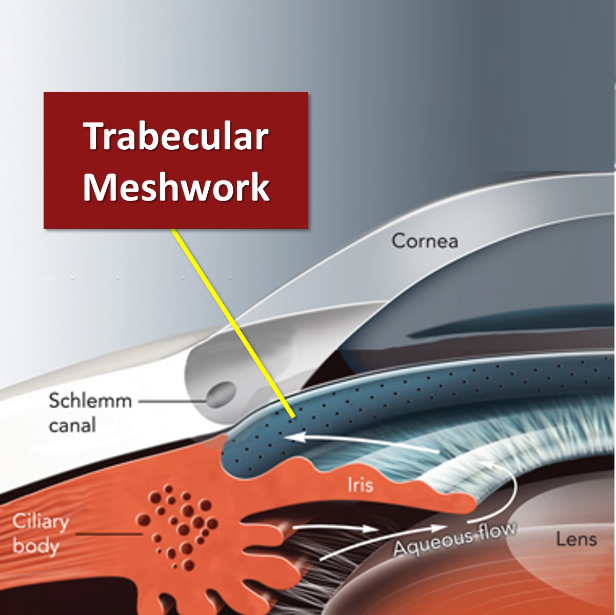
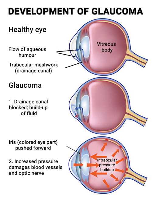
inbalance in aqueous humor production + drainage into the canal of Schlemm 📷 → incr. occular p → optic nerve death (ganglion nerve atrophy)
open angle:
- angle betw. iris + cornea normal
- decr. drainage due to:
- clogging + degeneration of trabecular mashwork - unknown cause
- unknown dysfunction in general
- chronic elevation in pressure → gradual vision loss (years)

closed angle:
- abrupt closure of iris + cornea angle
- rapid incr. of intraoccular p. → "acute glaucoma" → alert! 🚑
open angle → 80%
Open Angle Glaucoma
screening
because of the sneaky bastard character
- High intraocular pressure (obviously)
- Age
- Family history
- Race (African American and Hispanics)
- Suspicious optic nerve appearance (large vertical cupping)
- Thin central corneal thickness (** remember this one!)
PDP
- elevated intraocc. pressure >22mmHg (normal 10-22)
- optic disk changes (indicate ganglion atrophy) 📷
→ cup-to-disk-ration >0,5, or assymetry betw. eyes
→ abnormal ISNT-rule

- peripheral vision defect

corneal thickness
→ thick → incr. pressure
→ thin → decr. pressure
⇒ check with ultrasonic pachymeter


Acute Closed Angle Glaucoma
pupillary block:
lens plasters against back of iris → block aqueous flow through pupil → pressure gradient forces iris ant. →closure irido-corneal angle → blockage trabecular meshwork
- dark
- stressful situation
- drugs: antihistamined + cold meds
→ pupil dilation


acute:
- topical beta blocker (Timolol)
- CAIs (Diamox)
- i.v. osmotic agent (mannitol, glycerin) → draw fluid
- miotic agent (pilocarpine) → constrict pupil
- topical glycerin
to prevent recurrance:
- laser iridotomy 📷

Other types of glaucoma
- neovascular glaucoma
- pigment dispersion syndrome
- pseudoexfoliation syndrome
Neovascular glaucoma
retinal vein occulusion → VEGF (vascular endothelial growth fact) production → float through pupil → neovascularization iris → fibrous membrane in i-c angle → open-angle glaucoma → later: new vessel pull iris forward → closed angle glaucoma (irreversible)
- Laser peripheral retina → dec. VEGF production
- tube shunt
Pigment Dispersion Syndrome (PDS)
rubbing of pigmented surface of iris against zonules → shedding of some pigments → into aqu. humor → into trabcular meshwork +clogging
- high p after exercise
- pigments on gonioscopy
- trans-illumination defects on slitlamp
- Krukenberg spindle 📷

Pseudoexfoliation Syndrome (PXF)
BM-like material throughout the body + ant lens capsule → rough surface → iris rubbs off pigments through daily activity → clogging trabceular meshwork
Diabetic Retinopathy
glycosylation of vessel wall → over time: denaturation collagen → capillary thickening → breakdown
- signs of renal dmg: proteinuria, BUN+crea incr.
- duration of diabetes: after 10y →50%, 15y→90%
- good bloodsugar control
- avoid: smoking, HT, pregnancy
- NPDR: nonproliferative diabetic retinopathy

- PDR: Proliferative diabetic retinopathy
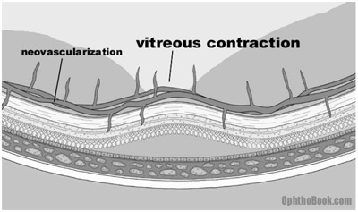
complete retinal vessel ischemia → VEGF → neovascularization → bypass the clogged artery → BUT arteries are abnormal in appearance + function (friable + leak) → also sprout into vitreos fluid → attach to vitreous proteins → when vitreos movement/contraction → pull on retina → retinal detachment + dmg to the friable arteries → intraoccular hemorrhage → vision loss → vessel scar down → massive traction on retina
→ can also lead to NVI (neovasc. of iris) → cover trabecular meshwork → neovascular glaucoma
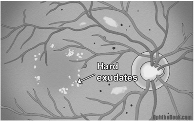
retinal macular edema
due to capillary+microaneurysm leakage
cloudy + elevated macula
yellow hard exudates (past edema) 📷

- Laser: seal off leaking vessels
- vitrectomy: removes humor + replaces with saline, and removal vitreos-retinal attachments


Retinal Detachments
separation sensory retina + underlying RPE (retinal pigment. epithelium) + chroroid plexus
→no further blood supply of the photoR → ischemia

macula
- myopic (near sighted) patients

- post surgery (catarct)
- traumatic sport injuries (boxing, football, and bungee-jumping)
📷 ←
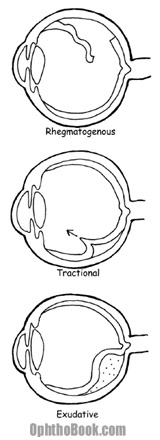
- rhegmatogenous (most common) → full thickness break (trauma, preexisting holes) →vitreous fluid into subretinal space → retina tear away + peel of underlying RPE + choroid → might spread along entire retina
- Traction: vitreous pulling or neovascular membrane contraction in diabetic retinopathy
- Exudative / Hemorrhagic: ocular tumor, inflammatory diseases, congenital abnormalities → breakdown of RPE+choroid plexus (blood-retina barrier) → pushing retina upwards
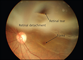
Retinal tear secondary to posterior vitreous detachment (PVD) 📷

- flashing lights (photopsias 📷) - like camera flash

- floaters 📷 - look like swarm of flies (blood cells or pigments in vitreous fluid)
- "dark curtain" 📷 - peripheral vision - most detachment involve periph. retina
REMEMBER: The combination of flashing lights and floaters should be considered a retinal detachment until proven otherwise.
- primary: surgery - esp. quick when macula is still attached
- laser therapy of sourrunding unattached retina
- cryo(probe) → also scarrs down retina by freezing from outside
- scleral buckling → silicon band as a belt → pushes RPE on retina
- pneumatic retinopexy → bubble of gas or silicon oil into eyeball → pushes retina back in place
- vitrectomy → retina manually floated back + also removal of scar tissue + neovascular membranes
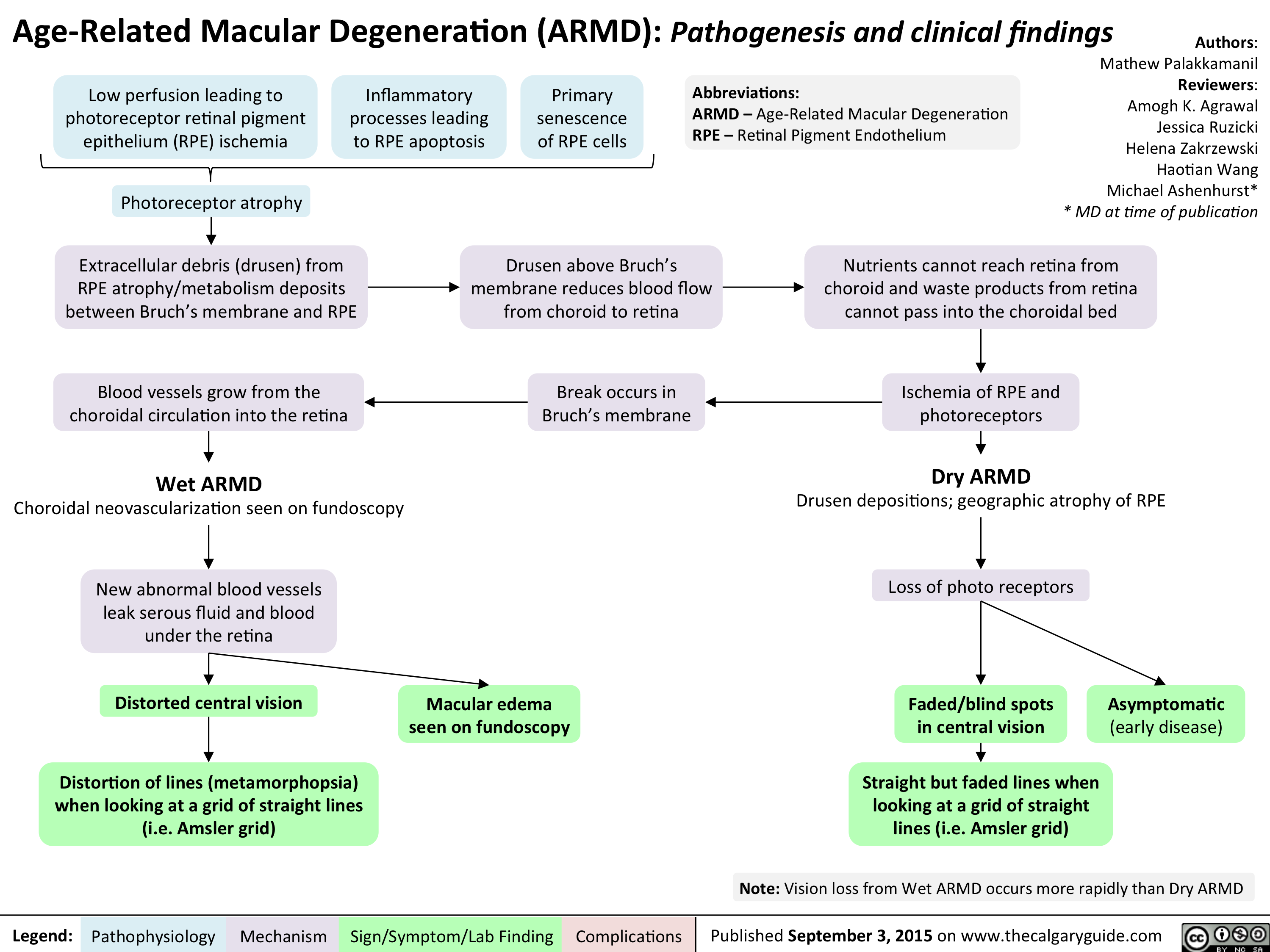
ARMD
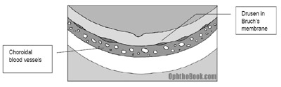
"drusen" (extracellular breakdown deposits) in Bruch's membrane which is betw. RPE/REtina + choroid → no nutrition + no removal of photoR waste products → retinal atrophy + pigmentary changes in macula → central poor vision
but slow progression (years)
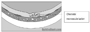
= neovascular ARMD
bruchs membrane break → vessel grow from chorid into retina → bleeding, edema, vision destruction
- PDT (photodynamic therapy) i.v. chemical that reacts to specific light wavelenght → focus light when in retinal blood vessel → coagulation
- Anti-VEGF drugs injection into eye → Avastin or Lucentis

Conjunctivitis
viral, bacterial, allergic
Viral (most common 50%)
- after upper resp infection by adenovirus
- red eye, watery tearing, NO discharge (little)
- uni or bilateral
- enlarged follicular bumps 📷
- swollen preauricular nodes
- enlarged follucular bumps inside of eyelid
symptomatic
Baterial
- pus (mucupurulent discharge)
- sticky eyelashes (due to creamy discharge) 📷
- papillary conjunctiva reaction
- unilateral
Etiologies:
- staph + strep,
- hemophilus influenza (in children)
- chlamydia + gonococcus (see below) → severe + sudden discharge
erythromycin
Allergic
- red, watery eyes
- itching + swelling
- allergic shiners (swelling around eyes)
- history of seasonal allergies
- concommitant allergic symtpoms: stuffy nose + cough
- bilateral
- antihistamines
- mast-cell stabilizers
- steroids
Blepharitis
stinging, tearing + "gritty" sensation (sand im auge)
- wash eyelashes with baby shampoo
- warm compresses
- topical ABs + steroids
- oral doxycycline → not AB-effect but changing fatty acid oil composition of meobomian glands
chalazion: noninfectious inflammatory granuloma rection
firm, mobile, nodular, nontender, nonpainful
- warm compresses, massage + lid scrubs
- incision + drainage
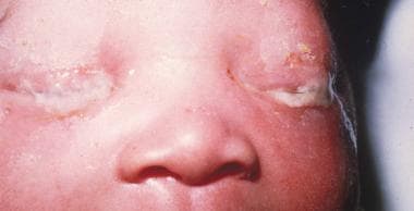
Chlamydial infection
Inclusion conjunctivitis aka "sexual" clamydial eyeinfection
- newborns (during delivery)
- horny teens with concurrent genital infection
- hand-eye
- shared cosmetics
- hottubs with not enough chlor
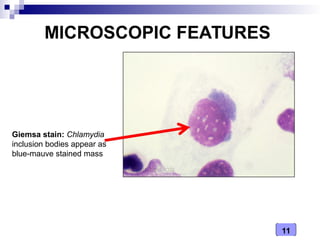
- immunofluorescence
- conjunctival culture → Giemsa → inclusion bodies 📷
- AB eyedrops + systemic azithromycin
- newborns → erythromycin

Trachoma aka "non-sexual" clamydial eye infection
- undeveloped countries with poor sanitation
→ leading cause of blindness in these countries

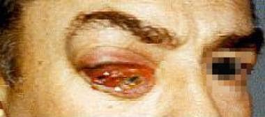
Gonococcal eye infection
very serious!
can penetrate cornea within 24-48h → endopthalmitis 📷 + loss of eye
- red
- severe + a lot of discharge
gram-negative diplococci
- ceftriaxone systemic!
- +topical AB
give prophylacticic erythromycin or silver nitrate(but gives "chemical conjunctivitis) after birth
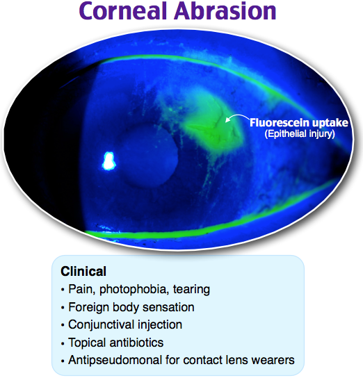
Corneal abrasions + ulcers
superficial epithelial defect
caused by trauma, infection, exposure
- severe pain
- intense photophobia
- "something is in the eye"
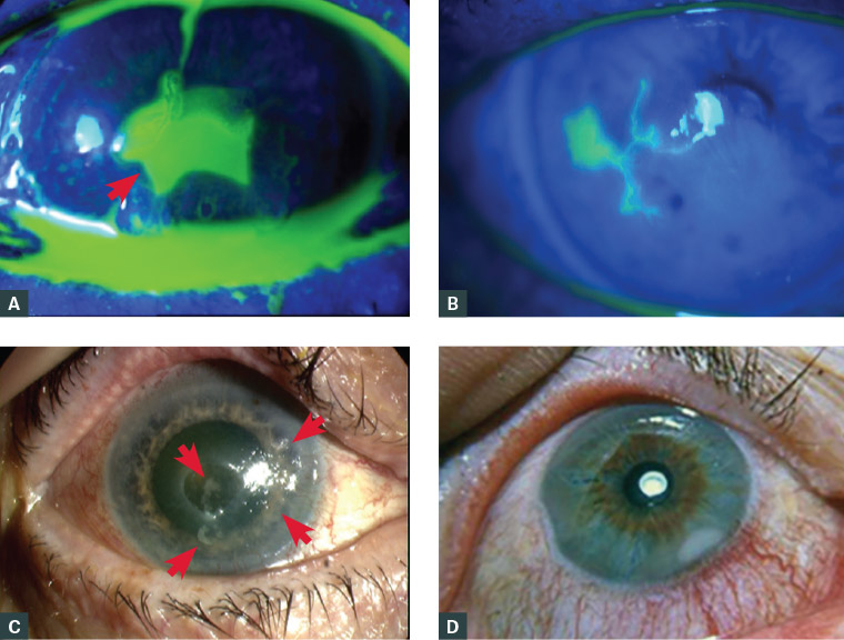
📷 flurosceine uptake → glow green under blue light
- aggressive lubrication → healing in 1-2days!
- empiric erythromycin
abrasion + associated bacterial infiltrate
aggressive AB aufn Nacken!
small → fluros (ciproflox)
large/central/not healing → culture → specific sensitive AB
Contact lenses
pseudomonas → ciproflox
caused by ext. foreign body → give more aggressive ABs
(e.g. tree branch, fingernal, soil)
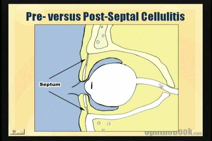
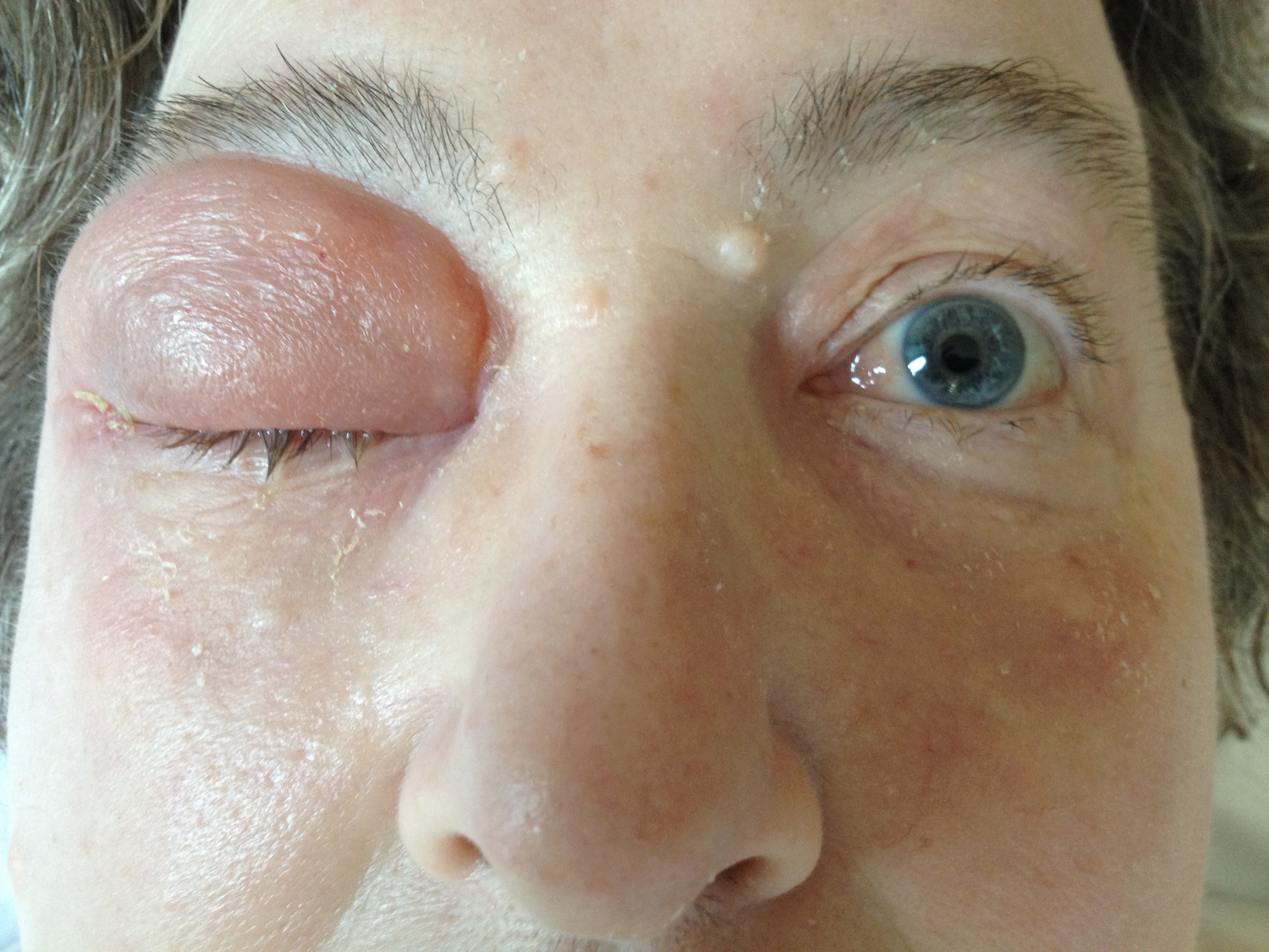
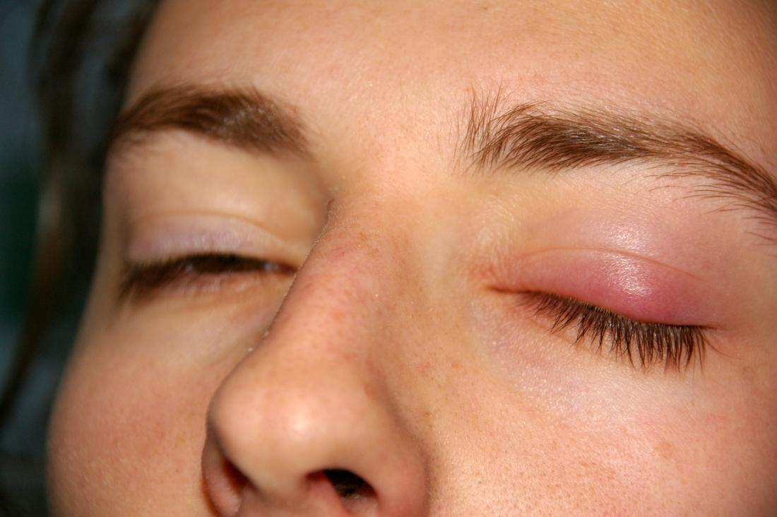
Pre- + post-septal cellulitis
signs of inflammation → swollen eyelid, erythema, warmth, fever
from tarsal plate to orbital rim 📷
post septal! = orbital cellulitis
superficial/preseptal looks bad but resolve without problem 📷
pics for orbital cellulitis later!
children → sinus disease
can also occur in patients with:
- tooth abscess
- fungal infection (immuno-compr)
- proptosis (exopthalmus)
- chemosis (swelling of conjunctiva) 📷
- decr. + painful eye motility (intraoc. muscle inflammation)
- decr. vision + RAPD(=marcus gunn pupil) (optic nerve dmg)
CT 📷
IV AB + surgical abscess drainage
Herpetic Keratitis
HSV type-1
mainly during childhood → latent: dormant in trigeminal ganglion → trigger (fever, trauma, psychological stress, UV sunlight)
initially only superficial cornea → heals + reactivate → deeper and deeper → corneal stroma scarring → sensory nerve death (decr. corneal sensitivity)

- Debridement (with cotton-tipped swab 📷)
- topical antiviral drops (Viroptic)
- Acyclovir orally

- corneal transplant (in severe corneal scarring)
- NSAID + mydriatic drops to prevent synnechia betw. lens + iris
AIDS Retinopathy
- cotton-wool (infarction of ganglion layer)
- microaneurysm
- hemorrhages
diabetes + HT
opportunistic infection with CMV (with CD4+ count <50)
→ necrotizing retinitis! 📷 → peripheral retinal necrosis (+ assoc. hemorrhages)
- antivirals: gancylclovir, foscarnet
but only virostatic → maintained treatment with gancyclovir or implant 📷
Endophthalmitis
infection inside the eyeball
- post surgery (even years)
- trauma
- endogenous infection spreads
often not the retina lel
filled with hazy inflammatory cells 📷
hypopyon 📷
pat. can see hand-motion or better → "tap and inject" = needle draw out sample culture + inject back broad-spectrum AB
if only light-perception or worse → vitrectomy (clean out the shit)
Diploplia
monooccular: refractive problem → most common astigmatism

binocular: eyes dont move synchron
→ CN3,4,6 palsy
→ extraoccular muscles abnormalities (muscle fibrosis in Graves)
→neuromuscular junction abnormalities (myasthenia gravis)
⇒ cover one eye → still double vision? → monoocular diplopia
Cranial Nerves and EOMs
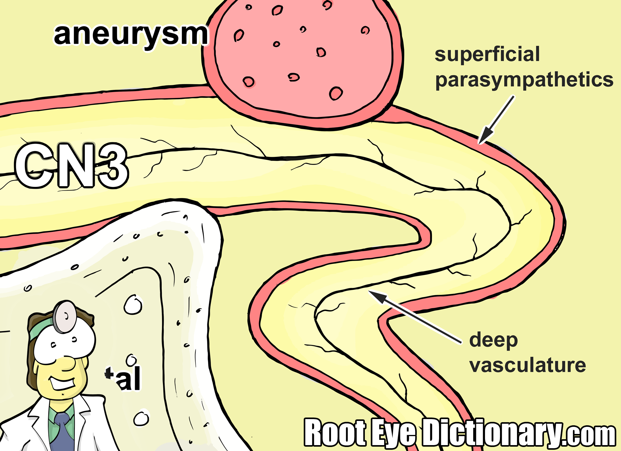
Occulomotor (III) Nerve Palsy
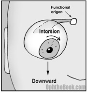
DOWN + OUT DEVIATION 📷
still functioning abducens → lateral rectus → pull lateral
still functioning trochlear → sup. oblique → depresses
PTOSIS → levatpr palpebrae dysfunction
"BLOWN PUPIL" (dilation) → parasympathic pupil constriction fibers travel within CN3
See also Neuromuscular anatomy
ischemic events due to DM or HT
compressive aneurysm pushing on nerve 📷
→ at junction of post. communicating artery + internal carotid artery
⇒BLOWN PUPIL
compressive lesion tend to involve pupil, vascular lesions spare it
MRI + angiography → rule out compressive cause (aneurysm or tumor)
blood pressure + gluc 🍨
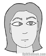
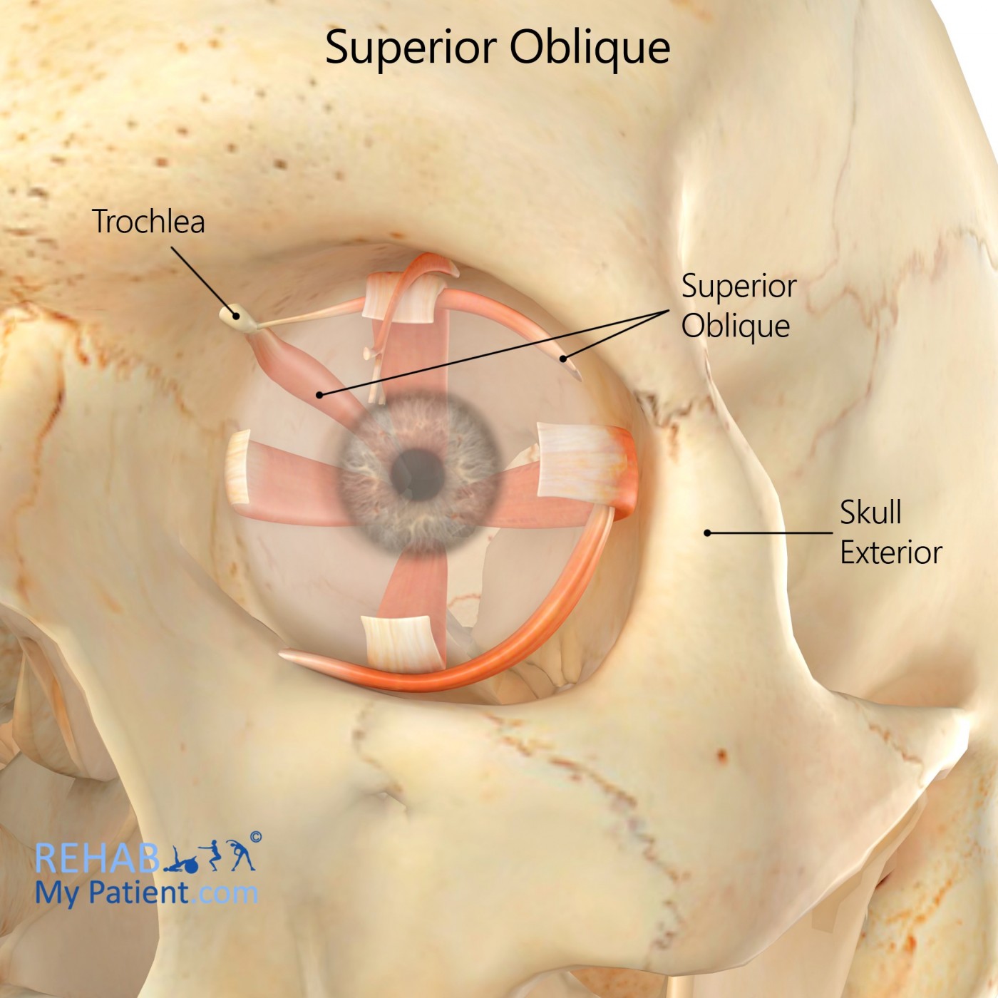
Trochlear (IV) Nerve Palsy
- upward deviation
- " cyclotorsion" twisting of the eye → tilt head away from lesion
normal action: 📷
- trauma
- congenital → check old pictures for head tilt → indicate an old/congenital palsy that has recently decompensated
- ischemic (diabetic, ht)
- tumor
Abducens (VI) Nerve Palsy
lateral rectus doesnt work → cross-eyed 📷
→ turn head to compensate
high ICP from pseudotumor cerebri

- CN3: The eyes are “down and out” with a droopy eyelid. Think of an aneurym if the pupil is blown.
- CN4: Patient tilts their head away from the lesion. Think of trauma or a congenital head-tilt that has decompensated with age. •
- CN6: The patient looks “cross-eyed.” Consider increased intracranial pressure.
Myasthenia Gravis
auto-antibodies against nictonic Acetylcholine receptors of striated muscles → block + destroy R
diploplia + ptosis
systemic:
- mastication problems
- drinking + swallowing problems
- talking problems
- pulmonary problems
when 30% of the R are destroyed
only striated
NOT smooth+cardiac


aspiration pneumonia + respiratory failure (inability to clear secretion)
Neuritis + Neuropathies of the Optic Nerve
Optic Neuritis
inflammation optic nerve → demyelination
associated with MS!

- decr. color vision
- pain with eye movement
- MRI enhancement of optic n.
- RAPD - marcus gunn pupil
- young patients
IV steroids

if brain involvement: enhancing lesions on MRI → interferons (avonex)
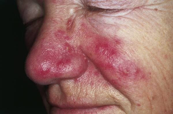
Giant cell arteritis - temporal arteritis
medium - small sized
around head - esp. temporal
- 60-80y
- sudden painless vision loss unilateral
- pathognomonic:
- scal tenderness + headache
- jaw claudication
- polymyalgias arm + shoulder
- B-symptomatic: fever, night sweat, weight loss
give high dose steroids asap!!
- ESP + CRP
- temporal artery biopsy → disruption internal elastic lamina giant cells
non-arteritic ischemic optic neuropathy → localized ischemia where nerve enters eyeball due to Arteriosclerosis
ESR + CRP normal
Pupillary abnormalities
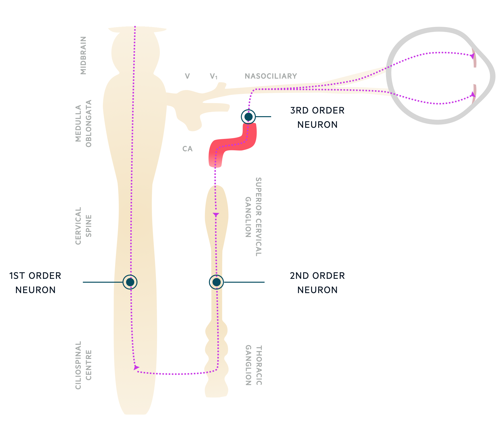
Horner Syndrome
sympathetic pathway which innervates the iris muscles gets knocked out
- ptosis
- meiosis (constriction) → can dilate at all
- scalp anhydrosis
Cocaine test → cocaina into eye → no dilation? → no sympathetic tone → horner sy.
Paradrine test: hydroxyamphetamine stimulates 3rd
→ still no dilation? → 3rd neuron is dead
→dilation → higher order problem!
- carotid dissection
- pancoast tumor
⇒ imaging!
carotid dissection! 🚑
Adie's Tonic Pupil
- opposite of horner sy
- dilated eye → doesnt constrict to light (but to near vision) 📷
- parasympathic plexus damage after viral infection
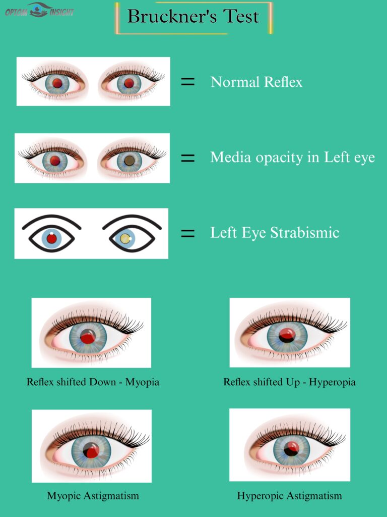
red reflex to figure out myopia/hyperopia (but not really accurate)
direct scope from distance
→ hows red reflex? 📷
myopia (inf. crescent)
hyperopia (sup. crescent)
hyperopia - small eyes + still growing
retinoscopy
flashing beam of light into eye → holding different power lenses in front → what focuses light properly + neutralizes red reflex
patch on good eye (without/with less refractory error or stabismus)
→ gives bad eye competitive advante to regrow afferent nerve fibers
DO IT ASAP! regrowth only possible until 7-10years
📷
- Esotropia - inward
- Exotropia - outward
- Esophoria - inward, only sometimes (stress, illness, fatique)
- Exophoria - outward, only sometimes (stress, illness, fatique)
actually a lot of children have some tropia which goes away in few month
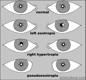
illusion of crossed eye due to nasal epicanthal skin folds → less sclera on nasal side
→ normal corneal-pupil light reflex 📷
Children unconciously supress their Strabismus to avoid seeing double which leads to amblyopia.
- non-surgical causes: correct refractive error + treat amblyopia → many cases will be resolved
- surgery: recessing medial or lateral rectus → reattach more post. → weakens the actions of this muscles and lets eye return in normal position
shaken baby syndrome
impressive retinal bleeding similar to that seen in Shaken Baby Syndrome (SBS) is only observed in cases of major trauma such as high-speed car accidents or falls from a significant height.
- Congenital cataract (idiopathic, genetic, metabolic(galactosemia), TORCH)
- Retinoblastoma (tumor of retinal photoreceptors → cream-colored mass filles eye with white creamy mass + cause reinal detachment)
- Retinopathy of prematurity 📷 (periph. retinal hasnt developed bloodvessel yet due to premature birth → ischemia → VEGF → neovascularization → bleeding + traction → retinal detachment ⇒compare diabetic retinopathy
Corneal Trauma
see corneal abrasion
Corneal laceration
!Perforation
Seidel test
wipe with fluorescin paper of wound → does it flow down the corneal surface? → seidel positive! ⇒ leaking aquaous fluid from the orbit washes it down
⇒ surgery
Orbital wall fractures
- vision (esp. color) → optic n.
- extraocular movement → entrapment?
- Proptosis (exophtalmus) or enopthalmus → use hertel exophtalmometer
- Palpation orbital rim
- Sensation of V1+V2 → forehead+cheek
- Keflex (cephalexin)
- Augmentin (Amoxi-Clavu)
- Afrin nasal spray (oxymetazolin) → decr. swelling of nasal mucosa
→ also no "nose blowing"!
Lid Laceration, Metal into eye + chemical injuries
how close is the laceration to the canicular (tear drainage) system?
→ put silicon tubes into nose to keep canaliculus patent before treating laceration 📷
- corneal abrasion → rust ring on cornea within a day
!if history metal-striking-metal → possibility of intraocular foreign body which entered the eye at highspeed and usually dont show really signs of injury → toxic to retina! → order CT and not MRI!
how quickly washed out
bases
Acids are less harmful than bases because acids have a tendency to cause precipitation of denatured proteins, which limits tissue damage. Bases, on the other hand, continue to cause damage without stopping, requiring continuous irrigation and pH monitoring until normalization.
Traumatic Iritis + Hyphema
cell + flare in ant. chamber

photophobia (inflammation → spasm of iris + ciliary bodies )
- topical steroids
- cycloplegic drugs (anti-muscarinic) → paralysis iris+ciliary muscles + dilation→ iris moves ant. + cant stick to lens and form synechia + helps photophobia (second. to spasm)
blood in anterior chamber (compare hypopyon)
often caused by blunt eye trauma
- steroids → encounter inflammatory response
- cyclopledic dilation drops → photophobia + prevent iris-lens-synechia
- glaucoma → blood clot clog trabecular meshwork
(+synechia betw. iris + lens)
Open Globe Injuries
nothing much about that stuff just dont push it because you might pop it like a grape
outcome is terrible + might need enucleation (raus mit dem apfel)
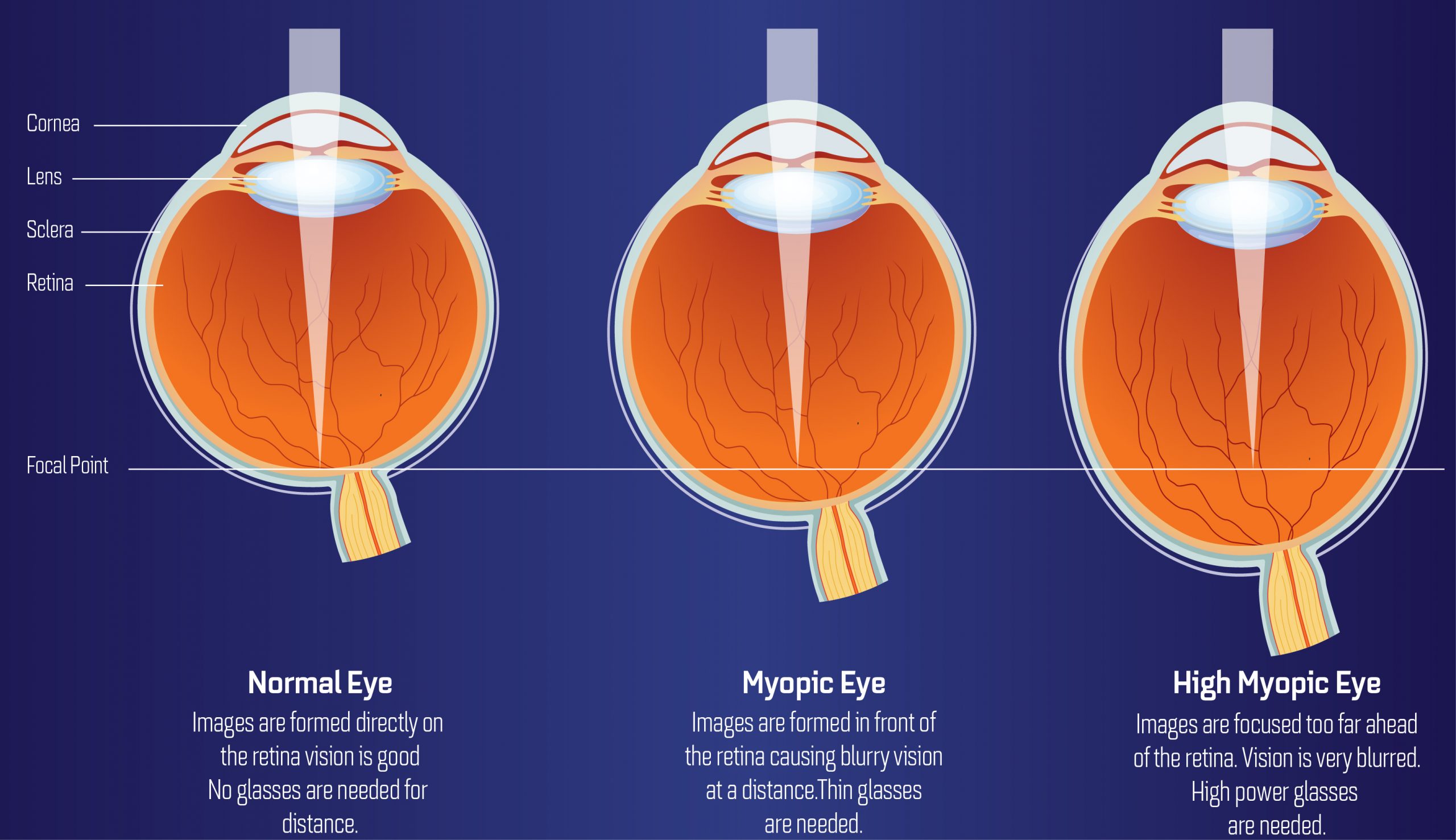
Myopia, Hyperopia and Presybyopia
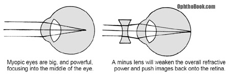
- nearsighted
- big + long eye
- light focus in front of retina
- give concave (minus lens) to weaken refractory power
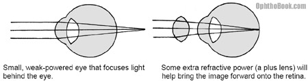
- farsighted
- small + short eye
- light focus behind eye
- convex (plus) lens to add refractory power
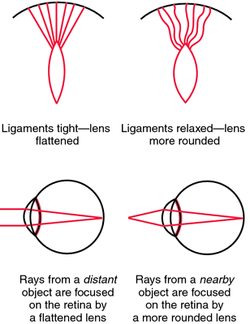
- minor to medium hyperopia are usually corrected by rounding of the lens (accomodation) and dont need any correction
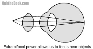
- age related less flexible lens (becomes dense + cant round out to accomodate)

- after 40
- corrected with bifocals
has part to correct nearsighted-ness + farsightedness
sphere
- in children
- difference of 2.00 diopters of both eyes →!might lead to amblyopia (one eye wont develop as good as the other) → plaster on the "better" eye needed
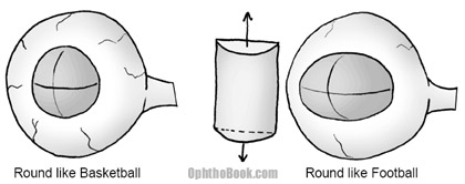
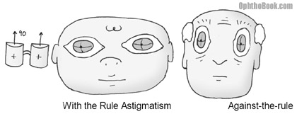
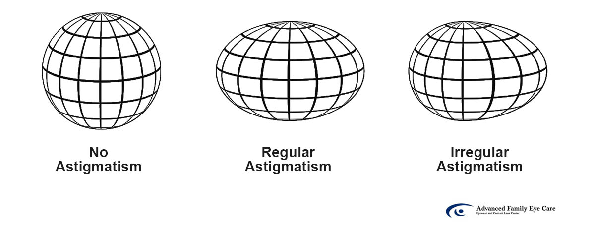
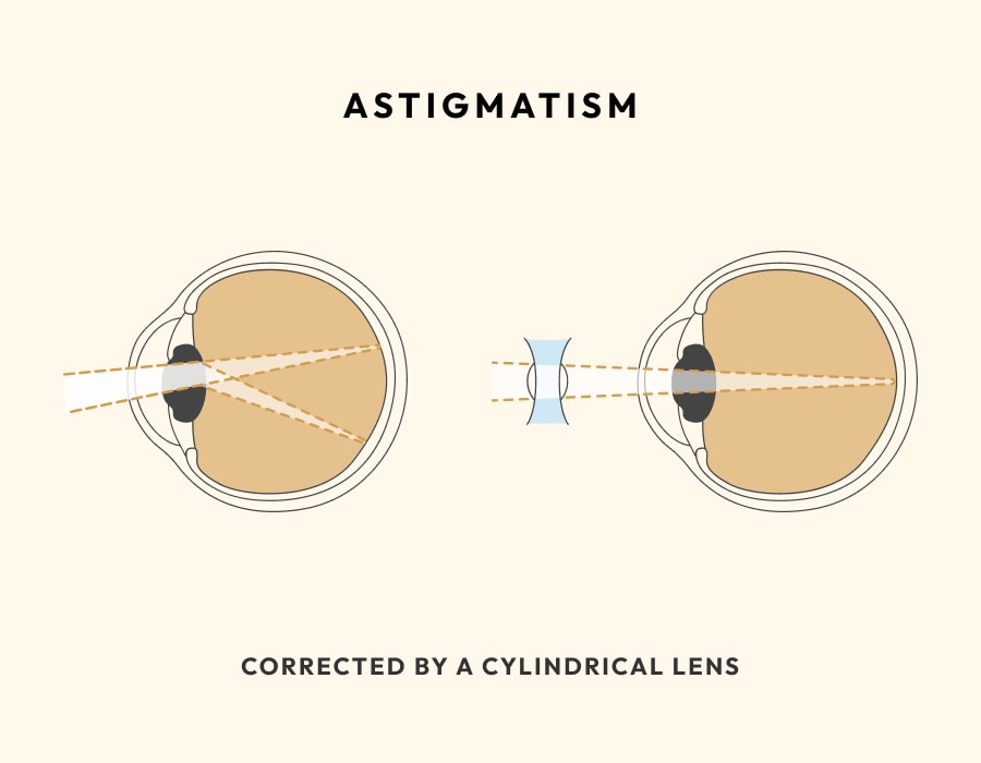
Astigmatism
device to determine glas prescription
has a big box full of different lenses that can be put in front of the eye
→ figure out spherical error, the extra cylinder to correct + angle of cylinder correction
lens is "footballshaped" in astigmatism and not "spherical" 📷
→ different refraction power at different points of the lens
cyclinder → how much cylindrical correction is needed to correct the refractory aberration along the axis? → add cylindrical lens
angle of cylinder 📷→ in what angle has the cylinder be placed to correct the "football" - if football is at 180° you need an angle of a cylinder of 90° (like in children)
foveal red-reflex
Basics
phakic - natural lens
pseudophakic - cataract replaces with artifical lens
aphakik - cataract removed but not replaced
⇒ 📷
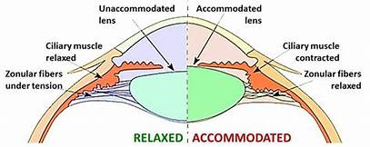
attached via zonular fibers to ciliary muscle
- controls lens focusing (accomodation-rounding by contraction)
- produce aqueous fluid
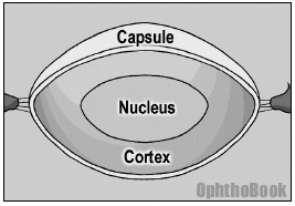
Cataract Types and Mechanism
nuclear sclerotic cataracts
over time lens (esp. nucleus) become larger and brunescent 📷(yellow or brown)
→ visual obstruction and problems with glare (blendung)
→ angle closure glaucoma (pushes iris forward)
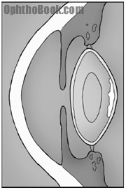
→ "Morgagnian cataract" 📷 : far advanced state → cortex liquefies+becomes milky and nucleus hardens+fall to the bottom of capsule
lens becomes round due to enlargement → correct coexisting presbyopia
post. subcapsular cataract 📷→ forms on post. capsule bag (looks like breadcrumbs or sand)
→ vision difficulty
- steroids use
- diabetes
- history of ocular inflammation
post → affects more the refractive power
- Posterior cataracts have a greater impact on visual complaints compared to anterior cataracts due to the optics of the eye.
- The eye has a refractive power of approximately 60 diopters, with 40 diopters from the cornea and 20 diopters from the lens.
- The nodal point of this system, located near the back of the lens, is crucial.
- Light rays are more affected the closer they are to this nodal point.
- Small posterior subcapsular (PSC) cataracts have more significance than larger anterior cataracts.
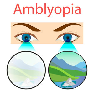
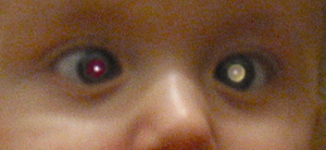
amblyopia 📷
other causes for leukocoria (📷 white pupillary reflex):
- TORCH infection

- retinoblastoma
- galactosemia
⇒ deadly!!
remove quickly within first 2 month → no implant because eye still grows → aphakic glasses/contacts → old enough: implant
trauma → capsule break → swells with water + turn white
CAVE! concommitant zonular tear
lens has no blood supply → nutrition due to glycolysis → high blood sugar → phosphorylation pathway (normal pathway) saturated → sorbitol pathway → sorbitol buildup in the lens → osmotic swelling → episodic blurred vision

appears after cataract surgery
residual lens epithelial left behind after surgery → migate along back of the implant → opcaify
YAG laser capsulotomy → little hole post. capsule→light can pass through
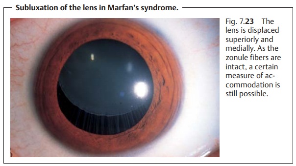
- trauma to zonules
- diseases affecting zonular strenghts:
- marfans → upward lens dislocation
- homocystinurea (zonules are composed of cysteine, without become fragile+break) → downward lens dislocation
Cataract surgery
- visual acuity (20/50=classic cutoff; but depends on pat. need)
- degress of glare
- underlying retinal diseases (DM); cataract interfere with fundus exam/laser treatment
- corneal curvature - the more powerful refraction of cornea, the less powerful lens - use keratometer to determine
- lenght of the eye - the short the more powerful lens - use A-scan mode ultrasound 📷
mnemoic: ASS EATING COCK PHAC (fuck) COCK INSERTION COCK POUNDING
- Anesthesia - topical tetracaine or retrobulbar block 📷
- Enter the eye - cutting cornea(fast) or tunneling sclera(slow but extendable if complication) 📷
- Capsulorhexis - hole into ant. lens 📷
- Phacoemulsification - phaco handpiece: oscillates - groove ridges into lens - broke into piece and eat up nucleus 📷
- Cortical removal - through suction (remove as much as possible→ see post. capsule opacification) → maintain post. capsule! 📷
- Insertion of the lens - foldable lens in remaining capsule (alternative if support is questionable: on capusle bag, on top of iris in ant. chamber, sutured to back of iris) 📷
- Close up - small incision=self-sealing; some require suturing
- Post-OP care: immediately antbiotic drops + shield over eye 📷; next day: AB drops + steroid drops


👁️ Eye anatomy & Visual function
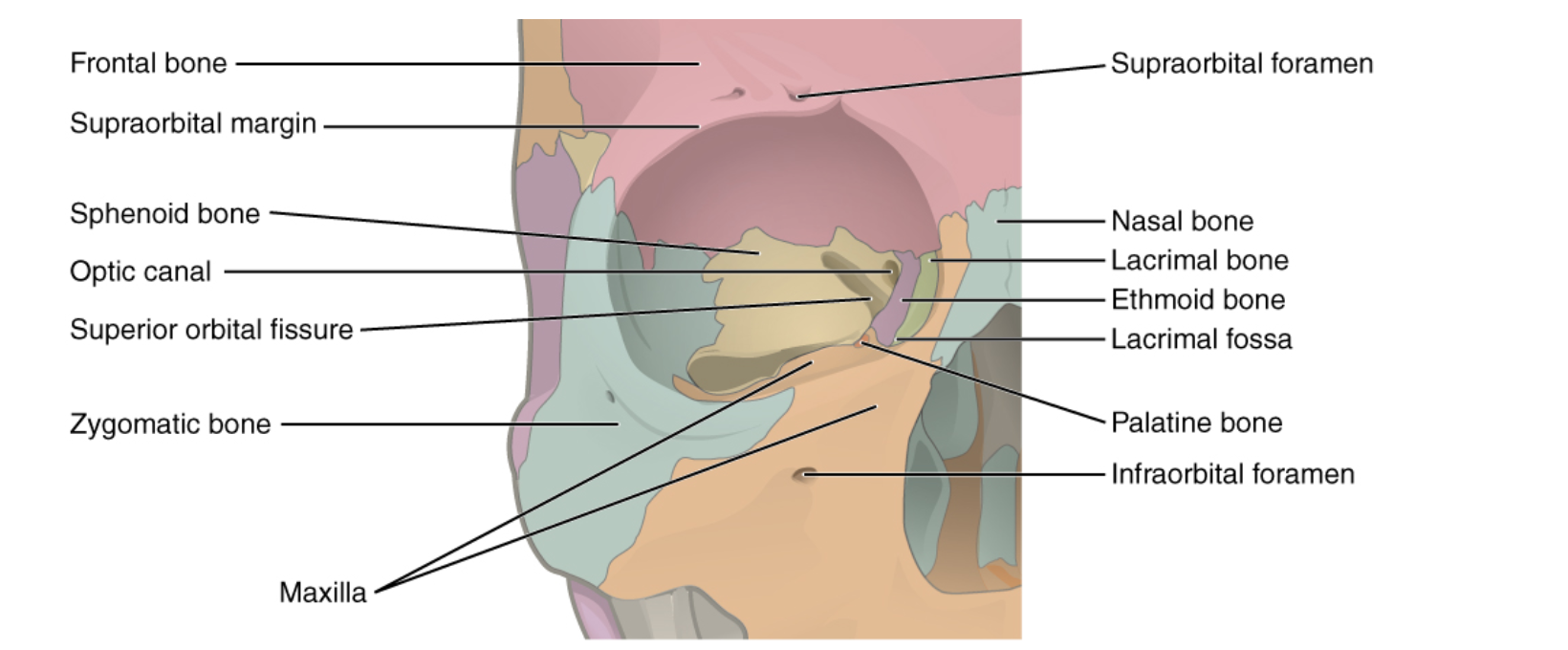
👁️ Anatomy of the eye
- sphenoid (lesser wing)
- frontal bone
ANATOMICAL PIC CAN BE FOUND IN THE END dont worry broddda
Trochlea 📷
- sphenoid (gr. wing)
- zygomatic bone
- zygomatic
- maxillary
- palatine
- maxillary
- lacrimal
- ethmoid bone
- sphenoid
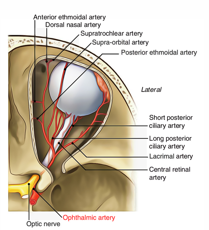
optic n + ophtalmic a.
- CN III, IV, VI
- V1 - opthalmic branch
- lacrimal a.
- ophthalmic v.
- globe
- lacrimal gland
- periocular muscles
- nerves
- orbital vessel
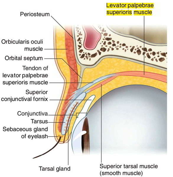
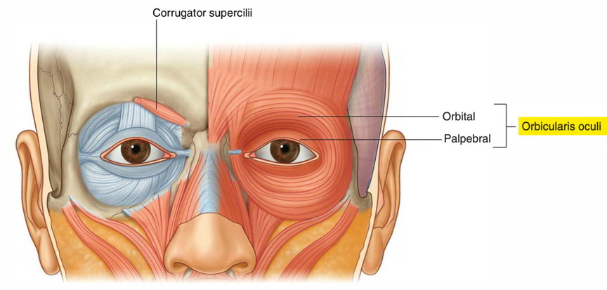
orbital + palpebral part
- pelpebral part consists of one or two lobule
- seperated by LPS muscle.
- Posteriorly-continuous with the orbital part
seperated by Levator palpebrae superioris tendon 📷
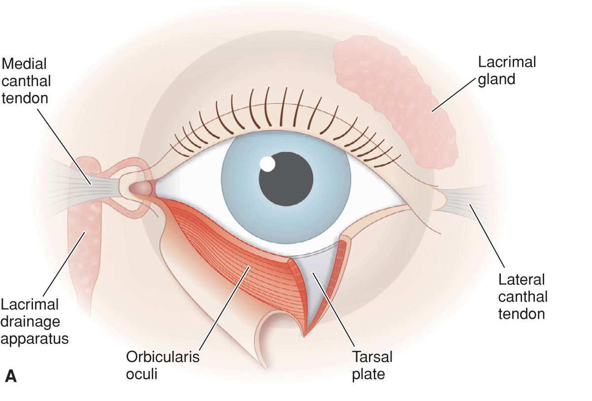
lacrimal puncta → lacrimal canaliculi → lacrimal sac → naso-lacrimal duct → inf. meatus
yes but:
balanced secretion - outflow
“Not all the tears get into the nasal cavity, a part of them evaporates. balance between secretion and outflow of tears so that a person does not realize he does have tears.”
- Rectus muscles: common tendinous ring (Zinn's tendon)
- Sup. oblique: sphenoid superiomedial to Zinn's tendon
- Inf. oblique: ant. maxilla, floor of orbit
- Levator palpebrea sup: sphenid bone
the “hook” to transmit the force of the sup. oblique m. → makes “rolling” movement of the eye possibe
CN IV trochlear- sup. oblique
CN VI abducens - lateral rectus
CN III occulomotor → rest:
- levator palpebrae superioris,
- superior rectus,
- medial rectus,
- inferior rectus
- inferior oblique
📷 orbicularis oculi - CN7
Buch und Präsi sind maximal belastend
TR:
protect + lubricate
tarsal plate = fibrous layers
- gives shape
- gives strenght
- muscle attachement
Meibomian glands
Secrete oil into tear film → keeps tearm from evaporating too quickly
Chalazion 📷 = ger: Hagelkorn
orbicularis oculi 📷
levator palpebrae
📷 “CN 3 opens the eye like a pillar, CN 7 closes like a fish-hook”
levator palpebrae - CN3
Orbicularis oculi - CN7
Tim Root:
no, transparent
no, starts at the limbus (see pic in Q before)
conjunctivitis
thickend part of the conjunctiva at medial canthus
Tim Root :
collagen
because the cornea relatively dehydrated
otherwise similar under microscope
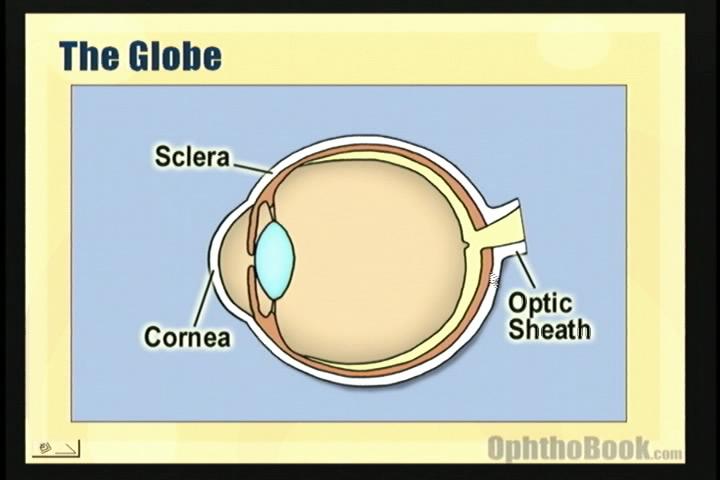
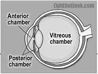
- Anterior chamber : betw. cornea-iris
- Posterior chamber: Iris + Lens
- Vitreous chamber: Lens-retina
2
vitreous humor + aqueous humor
- vitreous champer
- gel-suspension
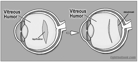
liquefy → PVD → might cause retinal tears
- With age and certain degenerative conditions, areas of the vitreous can liquefy.
- This can result in the vitreous falling in upon itself, known as a posterior vitreous detachment (PVD).
- While PVD is generally harmless, it can occasionally cause the vitreous to tug on the retina, leading to small retinal tears.
- Produced in post. chamber (ciliary body) → flows in ant. chamber → Drains back into venous circulation via canal of Schlemm
- High nutrient component → supplys avascular cornea + lens
glaucoma alert!
more infos later + check Tim Root
⇒ 📷
Tim Root:
Gives main refractive power (2/3)
- Outside → tears
- Inside → aqueous fluid
- Periphery → blood vessels
5 ⇒ 📷
nope, heals quickly but hurts as fuck
usually yes
Cant regenerate → remaining cells just get bigger → if to less cells → cornea swells with water
(physiological → pump function: water transport out of cornea)
Tim Root
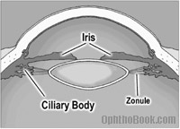
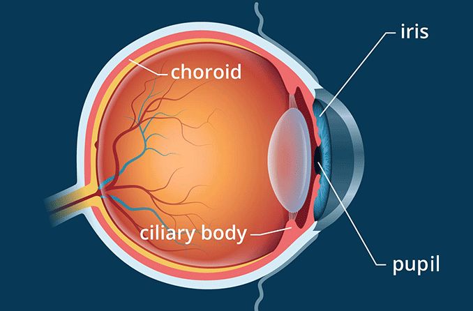
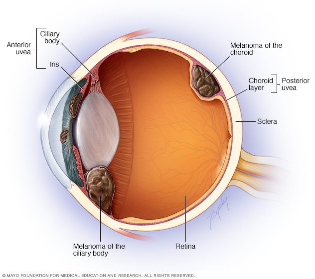
- iris + ciliary body + choroid plexus → continuity
- anterior uvea→ cilliary body and iris,
- posterior uvea→ choroid
spontaneously or with rheumatologic diseases
control amount of light hitting retina
- secretion aqueous fluid
- controls the shape of the lens → if contract → relax zonular fibers → round lens
bed of bloodvells → supplies outer 1/3 of retina → rods+cones
Retinal detachment → separation → RIP photoreceptors (die quick af)
Tim Root:
nope, get nourishe by aqueous fluid
highest in the body!! → 35% Protein, 65% water
zonules → suspensory ligaments
Presbyopia
Lens hardens → difficulties "rounding out" → can accomodate on near object
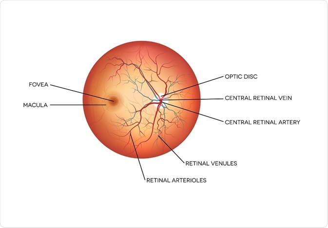
most inwards (towards vitreous)
pigmented area → responsible for central vision
fovea in the central part → extreme central vision
choroid
CAVE in retinal detachments
📷
contains ganglion nervs of optic nerve + retinal artery&vein ⇒ “cup”
🖼️ Visual function
- cones → iodopsine
- rodes → rhodopsine
Vit A
perception of light + reflection by visual cortex
light → decay of iodopsin → 1-6min
dark → regeneration of rhodopsin → 1hr
- How gucci is the retina?? = fundus exam/opthalmoscope
Difference of potential of the outer + inner layer of retina → different electrical response of retinal cells (photoR cells, bipolar cells,ganglion)
- ischemic events
- Diabetic retinopathy
- central retinal vein occulsuon
- Retinal detachments
- When the Fundus can't be visualized otherwise
- trauma
- vitreous hemorrhage
- dense cataracts
- Electrodes are typically placed on the cornea and the skin near the eye during an ERG recording. 📷
- ERG signals are small and measured in microvolts or nanovolts.
- ERG recordings involve exposing the patient's eyes to standardized stimuli.
- The resulting signal displays the time course of the signal's amplitude (voltage). 📷
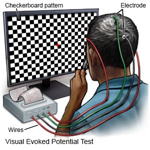
visual evoked potential 📷→ visual stimulus on computer screen → EEG (electrode on head) → how long does it take (time) for stimulus to reach occiptal cortex ⇒ check visual pathways (from retinalR → optic n. → visual cortex)
any signs + symptoms that could be indicative for a neurological cause:
- vision loss + alterations in color vision
- double or blurred vision
- flashing lights
- weakness eye,arm,legs (stroke)
sensitive in excluding lesion of optic n
R- lesion (but cant exclude optic n.)
optical pathway lesion
lecture says nictalopia = decr. VA+color during day + cone deficit (and hesperanopia is the thing mentioned below, but actually its the same thing 🖕🏽)
truth:
- night blindness
- congenital rod deciency
- or VitA deficiency
VA+VF
perception + reflection by the occipital cortex of the objects images formed on the retina
VA = perception of objects (by macular)
Minimum visible VA = stimulus appreciated by visual cortex as smth perceptual
Minum separable VA = discrimination 2 separate but adjacent stimuli
Snellen chart 📷
- refractory = myopia, stigmatism, etc. classic causes for glases
- opacification - caterac, corneal edema, vitrous hemorrhage, etc)
- optical n. + retinal illness: retinal detachment, diabetic retinop, ischemia, toxic, ...
- Amblyopia → without organic cause (strabismus)
bringt nüscht aka useless
-Thricromatic theory -Tetracromatic theory

tone (=color, 15 shades for 1tone)
saturation (how much radiation same wavelength)
brightness (how much white/cromatic color)
"Titten mit Sahne Bitte"
Congenital
- bilateral + symmetric
→ monochromatous → white + black
→ Dichromatous → no perception of one color
→ Trichromatous → normal
Gained
→ Chromatopsia → after cataract-surgery → blue vision for a while
→ cortical trouble
→ macular defects
🪞 Refractive errors
cornea
Ocular diopter (60D)
- cornea (45 D)
- lens (15D)
- statical → refraction without accomodation
- dynamic = accomodation → reflex induced by near-vision
Ametropias
→ Hyperopia
→ Myopia
→ Astigmatism
- image behind retina
- axial - short eyeball (90% of newborns)
- curvature → small curvature of cornea
types:
- low - up to + 2.0 D
- medium - up to + 5.0 D
- high - over + 5.0 D
minor to medium hyperopia are usually corrected by rounding of the lens (accomodation) and dont need any correction
components differentiated by eyedrops
- nonspecific, blurred vison,..
→ complications: strabismus(child), asthenopia, angle closure glaucoma
- convex lenses 📷 (glasses or contacts)
- Laser
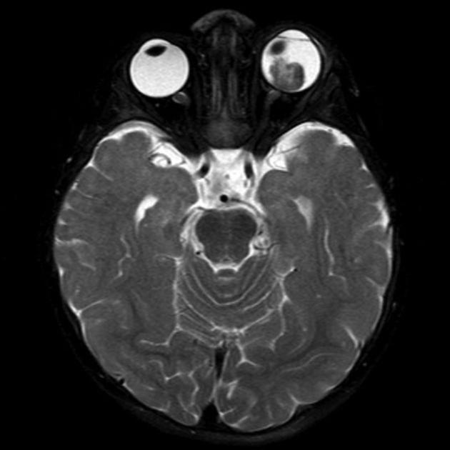
- image in front of retina
- axial - long eyeball
- curvature: high cornea curvature
- degree
- low (1-3)
- medium (4-6)
- high (>7)
- types
- simple → no retinal lesions + fundus
- progressive/malignant (>-10D) → lesions on fundus
blurred distance, squeezing eyes
think of big eye pulls everything
- cataract
- subluxated lens
- retinal detachment
glasses (concave), contacts, surgery (laser, new lens)
blurred, headache, night vision difficulty (seeing lights with a big star-like halo), eyestrain
The eye's ability to adjust its lens power to see clearly at different distances by modifying its curvature.
→ see TR (above)
- near point
- far point
- amplitude of accomodation (distance near + far p)
- asthenopia (ciliary m. fatiguabilitiy) - in hyperopia (+/- astigmatism) → high accomodation → blurred vision, pain,..
- Paralysis of accomodation (CN3 paralysis) - blurred near vision
- Spasm of accomodation (uncorrected refractive disorders, strabismus, drugs(morphine)) - blurred far vision
👓 Disorders of binocular vision
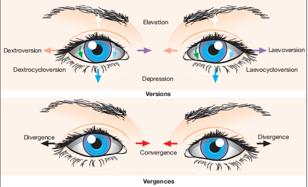
extrinsic eye muscle system 📷
versions: concomitant movements of both eyes in same direct
vergences: both eyes moves but opposite directions
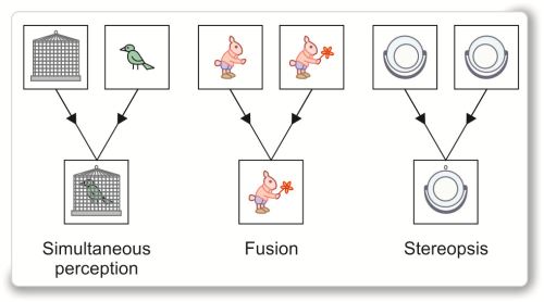
2 images on the 2 retinas → fusion in one sensation
SFS 📷
- Simultaneous projection: The ability of each eye to superimpose two different objects.
- Fusion: The ability of the eye to create a composite picture from two similar objects, differing in one detail.
- Stereopsis: The perception of the third dimension.
• Anatomical & optical integrity of the eye • The muscle integrity • Binocular visual field • Normal retinal correspondence (MRC)
normal retinal correspondence → corresponding points on the 2 retinas →esp. macula + fovea
children
squint = strabismus ⇒ double vision
paralytics → nerves palsy + neuromuscular
non paralytical → tropias + phorias
paralysis of ≥1 extrinsic occ. muscles
- congential
- Acquired
- inflammatory: encephalitis, MS, optic neuritis
- DM
- vascular lesions
- neoplastic regions
- trauma + degenerative
→ nerve dmg
- Diplopia
- False projection of object in space
- Nausea
Signs:
- squint
- cant move muscle in the direction of paralytical muscle
- different amount of eye devitation if you compare both eye
- torticolis → correction of diploplia (compare CN4 palsy)
- Diplopia early
- homonimous - abductor paralysis
- heteronimous - adductor paralysis
- False projectior - an anormal appreciation of an object in space
- Nausea
paralytical features that are not present in non-paralytical:

- cant move eye in the direction of paralytical muscle
- different amount of eye devitation if you compare both eye
- diplopia
Heterophoria is an ocular condition where there is a latent misalignment of the eyes when they are at rest, rather than actively focusing on an object. It can lead to eye strain but doesn't typically result in double vision or other noticeable symptoms.
caused by uncorrected refraction error
- headache
- pain
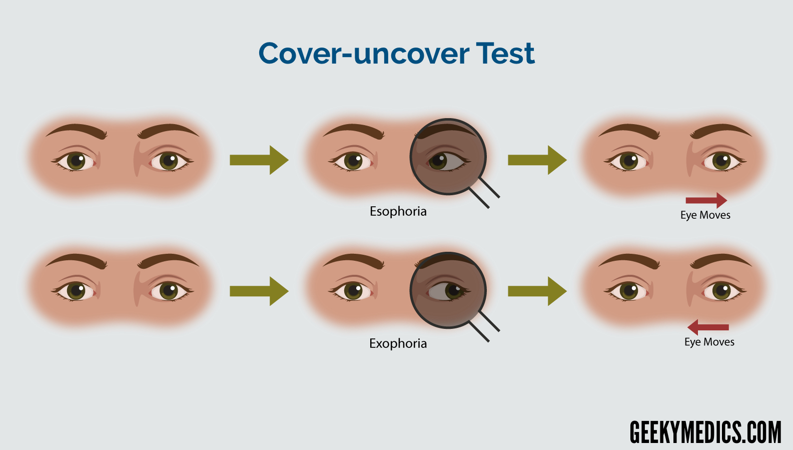
- cover-uncover test +
- angle constant in all direction of gaze, eye movement normal, deviatian is interemittent
cover-uncover test = presence of squint? → pat. fixate small target, cover one eye (2-3sec), quickly remove, covered eye deviated is delayed 📷
- optical correct
- synptofore treatment 📷
- refractory defects (hyperopia, myopia,astigmatism)
- sensorial defect (leucocoric pupil= cong. cataracts, TORcH, retinoblastoma)
- anatomical + motor defect
direction, onset, location
- direction (eso, exotropia)
- onset (congenital<8m, late>2y)
- location (monolateral, bilat)
Esotropia
- Congenital
- Late onset
Exotropia
- Divergence excess
- Convergence insufficiency
→📷
- VA decr. in monolat. squint → ambylopia
- constant squint
- cover test (covering fixing eye, deviated eye needs to move to fixate) 📷
- mobility normal (DD paralytical)
- optical correction of refractory errors
- treat ambylopia (cover with plaster 📷)
- orthoptic → synoptophore (for low amplitude - such rather used in -phorias)
- prism
- surgery (when everything else doesnt work → resection of medial/lat. rectus) → evtl. orthoptic postsurgery
🏐 Pathology of the orbit
Watch Tim Root - Eye infections
ant. (=preseptal cellulitis) + post
ST-infection ant. to orbital septum due to
- skin trauma
- spread of infection
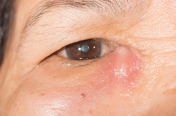
- acute hordeolum(inflammation of zeis gland 📷)
- dacryocystitis (lacrimal sac)
- upper resp tract or middle ear
- unilateral
- tenderness
- periorbital redness
- lid swelling
- minimum proptosis (exophthalmus)
- VA + occular motility normal (DD!)
systemic AB (penicillin/oxacllin)
into posterior osteoperiositis
same but additionally
sphenoidal aperature syndrome (CN3,4,6 + branches of CN V1 passing through sup. orbital fissure 📷)
- proptosis
- immobility of the globe
- anesthesia (CN V impairment)
orbital peak syndrome
- ↓ of VA (compression Optic n.)
Evolution: Orbital cellulitis
Tx: Systemic ABs + Etiological
infection of the soft tissues behind the orbital septum 📷
Staph. Aureus/Strep/H. influenza:
- spread from sinus disease
- spread from ant cellulitis
- spread from dacryocysititis
- posttraumatic
- post-surgery
- Inflammatory/Infectious signs: generel sympt + signs of infection + inflammation (malaise, fever, pain, swollen, red, warm, tender)
- moderate proptosis (exophthalmus)
- painful ocular movement
- + all of the others
- Orbital abcess
- Cavernous sinus thrombosis +Central retinal artery/vein occlusion
- Optic neuritis + intracranial involvement
- exposure keratopathy (due to huge proptosis cant close both eyelids → exposure of inf. cornea all the time)
- AB! (cephalo, metronidazole; Ampi+Pen(children))
- abcess → incision +drainage
= complication of orbital cellulitis
AB + anticoagulation
inflammation of the extraocular muscle
(usually idiopathic)
chronic = less pain
→ steroids + radiotherapy!
radiotherapy might produce cateracs → not really used anymore
- proptosis
- painless, irreductible
- if behind muscle cone → decr. VA, retinal hemorrhage + dilated retinal veins
- if inside muscle cone → diploplia + extraoccular muscle paralysis
imaging! location + extension
CT 📷, MRI (xray)
- primary
- secondary - from NS, sinus, occular globe
- metastatic
classics
- Surgical
- radiotherapy
- chemo
🥠 Eyelid pathology
almost always unilateral 📷
ant. uveitis → give mydriatic eyedrops + NSAID to prevent synnechia development (!closed angle glaucoma)
- after trauma
- pyogenes or StaphAureus
inflamm + pus
- pain
- lid edema + erythema
- suppuration of eyelid (localized [i.e. hordelum] or diffuse)
- systemic : Penicillin
- surgical : incision + drainage
ac. staphylococcal abscess of zeiss + lash follicle 📷
- non-pharma: hot compresses + epilation of eyelash
- local AB ointments 3x/day
= small abcess of Meibomian glands
- tender
- swelling within the tarsal plate
- local AB ointment
- surgical
meibomian cyst a chronic lipogranulomatous inflammatory lesion caused by blockage of gland orifices and stagnation of sebaceous secretions.
increase risk for chalazion for pat with acnea rosacea or seborrheic dermatitis
onset: painless nodule
- occasionally upper lid chalazion may press on the cornea and cause blurred vision ( induced astigmatism)
- painless, roundish, firm lesion in the tarsal plate.
- eversion of the lid may show associated polypoid granuloma (if lesion has ruptured through the tarsal conjunctiva.)
- +- associated blepharitis or acnea rosacea.
- small chalazia may disappear spontaneoulsly,
- persistent lesions ⇒ surgical treatment.
inflammation lid margin by staph aureus
- Erythematous → hyperemia
- Seborheic - crusts (hard+brittle) base of eyelash 📷
- Necrotic (rare)→ intrafollicular abcess, scars, necrosis, absence of eyelashes
inflammation of sourrounding:
- ext. hordeolum
- conjunctivitis
- marginal keratitis 📷
- topical ABs + steroids
- wash eyelashes with baby shampoo
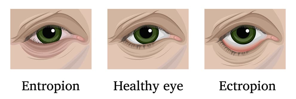
lower eylids turned outward 📷
- asympt
- tearing / irritation
typically senile
paralytical (VII palsy)
scars (chemical burn)
Types:
1.congenital 2. acquired - involutional (senile)
surgery
grafts, suturing (tarsorrhaphy for paralytic)
- cicatriceal - chemical burnsurgical (lid grafts) tarsorrhaphy
- paralytic: CN VII palsy
tarsorrhaphy suturing of the eyelids
=lid margin is turned inward the globe ulceration of the cornea
senile
scars→ after conjunctivitis (i.e. clamydial conj)
spastic - only intermittent
Types : 1. congenital 2. acquired
- involutional (senile)
- cicatriceal
- spastic
Schnippschnapp surgery
= involuntary closure of the eyelid 📷
unknown etiology
- incontrolled blinking, twiching or closure of the eyelids
- involuntary contractions of the orbicularis oculi muscle
- botulinum toxin injection in orbiculari muscle
- surgical (excision of the orbicularis muscle of upper lid)
lid margin is abnormally low (malposition) → insufficient upper eyelid retraction
- congenital
- senile
- myogenic (myasthenia gravis)
- neurogenic (CN3)
- traumitc
- tumor
surgery
☄️ Conjunctiva pathology
- staph aureus
- h. influenca
classic
watery discharge, red
morax axenfeld diplobacillus
- itching
- redness at lat+ medial canthus 📷
Zinc sulphate

- low grade inflammation of lid margin
- colonization of meibomian gland
during delivery from mother
3-5days after birth
- Bilateral
- discharge → tightly closed eyes due to stickyness
- classic conj. symptoms (red, swollen, chemosis)
corneal ulcer → corneal perforation → lens damage, endophtalmitis (inflamm. of eyeball), scar formation
crede method (silver nitrate) drops after birth
or erythmycin
Topical & Systemic Penicillin (cefrotriax in adults)
gonorrhea
basically same as neonatal BUT UNILATERAL
- male patient
- onset - 3-5 days of incubation
- unilat
- purulent discharge
- conjunctiva-congestion+ chemosis
penicillin or ceftriax systemic
topical: macrolides
children, young active teens
Chlamidia oculogenitalis 👁️🍆
5-19days after birth
- Incubation period: 5-19 days after birth (📌 notice: later than gonorrhea!)
- Symptoms: red and swollen eyelids, purulent discharge 📷
topical AB (+ macrolide systemically)
The clinical features and the treatment are the same as in inclusion conjunctivitis
→ Staph, Strep, pneumococcus
Corynebact. diphteriae
pseudomembranes 📷→ removal of the membranes → does NOT lead to hemorrhage
- symblepharon (scarring)📷
- eyelid deformation (entropion/ectropion)
- diphteria anti-toxin
- topical AB
in specific regions (endemic)
low hygiene
Chlamidia trachomatis → patient in childhood
- male + young (adults,children)
- recurrent bilat.
- allergic mechanism (eosinophil mediated)→ Atopy: allergic sympt (rhinitis + asthma)

- ocular itching
- mucus discharge
steroids → topical + systemic
- tuberculoprot. or toxic agent(sepsis)→ allergic reaction to foreign antigen (nowadays most commonly due to Staphylococcus ihr wichser!)
- young children

- etiological (tbc)
- Steroids + atropine
💧 Lacrimal pathology
inflammation lacrimal gland
- sinus infection (acute)
- Tbc, syphilis (chronic)
- acute discomfort + swelling, tenderness
- S-shaped ptosis
- reduction in tear secretion as compared with the other side
lacrimal gland tumor - no inflammatory signs
chalazion - no horizontal s-signs
NSAID + AB
inflammation lacrimal sac → obstruction of nasolacrimal duct
- idiopathic or
- aquired:
- idiopathic stenosis
- trauma
- tumor
- epiphora (excessive tears)
- pressing on lacrimal sac → reflux purulent material from lacrimal punctum 📷
no red eye → DD: purulent conjunctivits
- massage of the lacrimal sac
- nasal probing 📷
acute + chronic
- medial cantus sudden pain+tender!! + signs of inflammation
- epiphora
same as congenital AND painless swelling
- epiphora
- painless swelling at the inner canthus
- pressure on the sac → reflux mucopurulent material
- systemic AB
- dacryocystorinostomy
🦏 Cornea pathology
- Classification:
- Ethiological: microbial, viral, fungal
- Pathogenetic:
- Exogenous: ocular trauma, corneal ulcer
- Endogenous: endotoxins (specifically Gram-negative bacteria induce inflammation)
- Location:
- Superficial keratitis
- Stromal keratitis (interstitial)
- PAIN
- PHOTOPHOBIA!
- decr. VA
- Superficial, suppurative keratitis
- caused by:
- pneumococcus
- staphylococcus
- Pseudomonas
- i.e. due to contact lenses, poor hygiene
- chronic dacryocystits
- underlying corneal disease
- Dry eyes
- Contact lenses
- Topical/ systemic Immunosupressive agents
extension descemet membrane → descematocele→ corneal perforation → lens-iris-synechia → intereference with aquaous humor flow → second. closed angle glaucoma
that shit is serious → keep in hospital
- AB
- topical Ciproflox
- p.b. = peribulbar Gentamicin
- systemic Cephalos
- topical cycloplegics: Atropine
- if secondary glaucoma → anti-glaucoma drugs
- if perforation → therapeutical contacts, corneal grafts
- primary → childhood → self-limited
- Gasser Ganglion (Trigeminal nerve ganglion) 📷→ reactivation → cornea
- photophobia
- dendtritic ulcer (fluoresceine) 📷
- stromal infiltrates
- decr. corneal sensitivity
- decr. visual aquity
- foreign body sensation
- peri-limbical congestion
- Recurrences and progression to disciform keratitis are observed in some cases.
- Improvement in writing and formatting of the marked text has been made. The language tone has been adjusted to be confident and academic while maintaining the medical terminology.
- acyclovir drops (topical + systemic)
- NSAID
- debridement
- bandage lens 📷
bandage contact lens is designed to protect an injured or diseased cornea from the mechanical rubbing of the blinking eyelids, therefore allowing it to heal. The Bandage lens often makes the eye feel more comfortable. It is usually a soft lens, but not always.
follows superficial herpetic Keratitis
- antiviral (topic + syst) → Acyclovir
- cyclopledigic (Atropine), topical steroids + systemic NSAID
H. zoster
elderly + bad immunity (AIDS, diabetes)
like HSV
antivirals + bandage lens+ debridement
→ immunity → no recurrence
(Tx is the same)
- Congenital syphilis
- Tuberculosis (TB)
- Leprosy
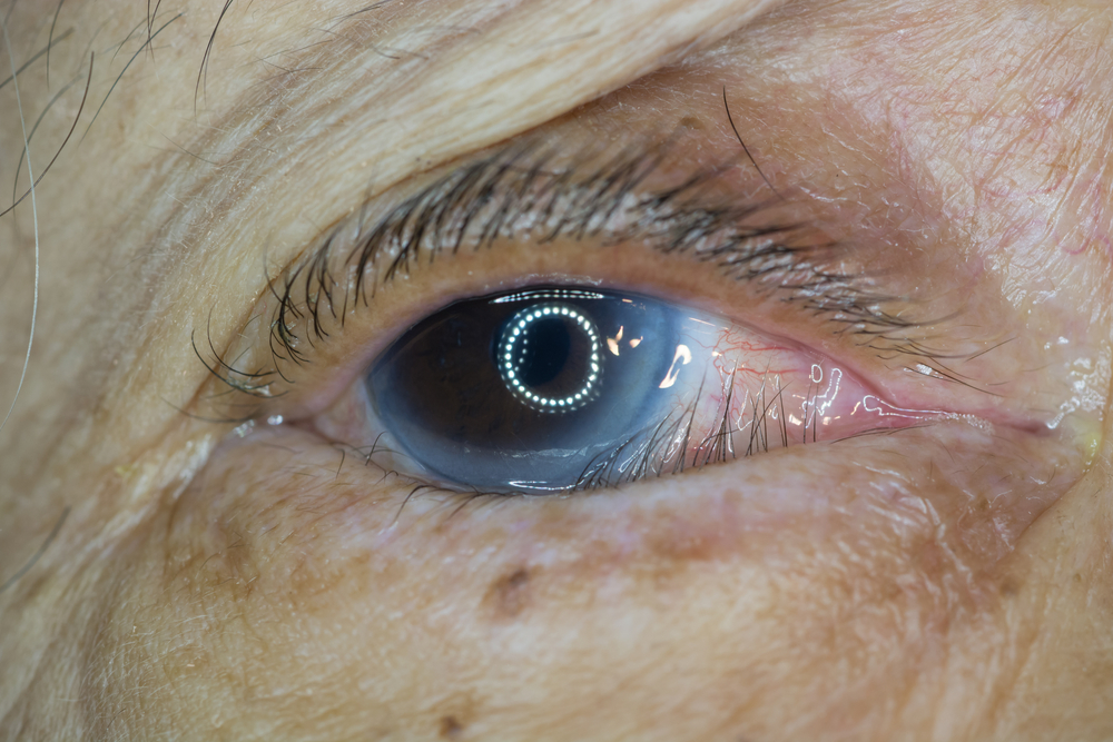
obstructed vessels → after a period of time new vessels form ⇒ fine thin vessels in corneal stroma = Ghostvessels 📷 (→ retrospective dg)
- The cornea exhibits diffuse stromal haze.
- High magnification reveals ghost (empty) vessels in the deep and mid-stroma.
- The cornea may have reduced thickness.
- This condition typically affects both eyes.
- etiology
- steroids + atropine → topical
- penicillin systemically (syphilis)
poor contact lens hygiene/extended wearing
(acanthomoabae)
- ocular pain, decr. VA
- Slit lamp: paracentral ring infiltrate 📷
- antifungal drops + neomycine (aminoglycoside) drops
- surgery →quick evolution to corneal performation! → penetrating keratoplasty 📷
→ improper cornea weting, lack of palpebral occulusion
CN7 palsy
proptosis
ectropion

- corneal erosions → ulcer
- neovascularization
- Etiological
- Artificial tears
- Soft bandage contact lens
- Surgical
- congenital
- acquired
- injury CNV + other causes → anaestetic cornea → exfoliation of epithelial cells + edema (corneal dystrophy)
- Viral: HSV, Zona zoster
- DM
- punctate erosions → ulceration
- gray epithelial cells
- surgery 📷
- artificial tears
- treatment of ulcer
hereditary genetic bruder
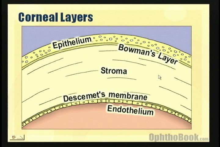
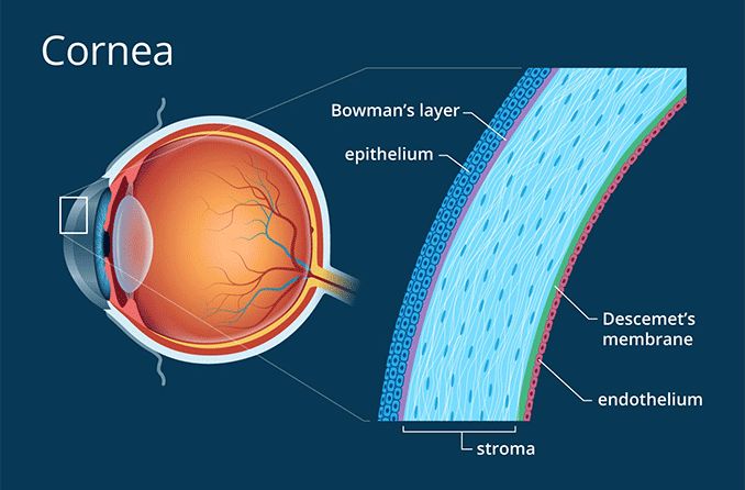
epithelium, stroma, edothelium 📷
🌈 Uveal pathology
- exogenous
- septic (direct introduction due to OP or perforation)
- toxic (toxine introduction due to ulcers + chemical burns)
- endogenous
- infections
- Rheumatism
- Sarcoidosis
- Collagen
- pain
- photophobia
- synechias → glaucoma
- flare + cells in ant. chamber (Tyndal +)
- decr. VA
- red eye
- gray iris
- panuvitis
- secondary closed angle glaucoma
- complicated cataract (because of inflammation + steroids use)
- atrophy eyeball
- AB + NSAID
- steroids
- cyclopledic drugs
- beta blockers (in glaucoma)
- surgery (in cataract)
- etiologic spefic
perforation ocular wound by staph or pseudomonas
- ocular pain
- decr. VA
- hypopion + post. synechia
- pupillar exudates
- edema of wound + lid
panopthalmitis → poor prognosis

- AB
- steroids
- cyclopledic drugs
- NSAID
- removal of the eye (Evisceration 📷)
spread of endogenous infection/inflammation:
atypical agents
- toxoplasmosis
- TBC, syphilis
- sarcoidosis
- minigits, endocarditis...
- dec. VA
- vitreos exudate
- choroid edema + nodules
- cataract
- neuroretinitis (due to spread)
- AB
- steroids
- NSAID
- perforating wound or ulcer
- foreign body retained in eye
- intraoccular tumor
idiopathic
viral infection + associated allergic reacting in the injured + non-injured eye → uveitis in non-injured eye after a period of time (evtl. after years)
- correctly suture the injured eye
- radical removal of the injured eye
- There is no known benefit to removing the exciting eye after the onset of sympathetic ophthalmia.
steroids, steroids, steroids
🥚 Lens pathology
Watch Tim Root: Lens + Cataract surgery
amblyopia 📷
- Congenital cataract (idiopathic, genetic, metabolic(galactosemia), TORCH*)
- Retinoblastoma (tumor of retinal photoreceptors → cream-colored mass filles eye with white creamy mass + cause reinal detachment)
- Retinopathy of prematurity 📷(periph. retinal hasnt developed bloodvessel yet due to premature birth → ischemia → VEGF → neovascularization → bleeding + traction → retinal detachment ⇒compare diabetic retinopathy

remove quickly within first 2 month → no implant because eye still grows → aphakic glasses/contacts → old enough: implant
cortical, nuclear, post. subcapsular cataract
- Incipient:
- accumulation of liquid betw. lens fibers
- Immature (intumescent):
- incr. fluid → swelling → glaucoma (closed-angle)
- decr. VA
- Mature
- complete opacification
- very low VA
- Hypermature
- liquefaction cortex, zonules, ant. capsule
- open-angle glaucoma (→leakage of lens material into ant. chamber through defective capsule→ phagocytes → blockage of trabecular meshwork)
→ nucleus sinks to lower part
- nucleus opacification
- post. subcapsular cataract → decr. VA from the beginning!
other occluar diseases
- ant. inflammatory process → reccurent uveitis, keratitis
- high myopia
- glaucoma treatment
- retinitis pigmentosa (often cause subcapsular c.)


caused by general diseases / drugs
- DM
- scleroderma
- corticosteroids (systemic + topical)
→ steroids + DM causes esp. post subcapsular c.

see TR - Post Capsular Opacification
marfan
homocystinura
trauma
Hypermature cataract
incomplete dislocation (sublux) of the lens
aphakic zone + lens zone
glaucoma
anterior → lens into AC posterior → lens on Retina
- pain + decr. VA
- decr. depth of ant chamber
- lens in ant. chamber
- second. glaucoma
- corneal degeneration
- anterior uveitis
- low VA
- deep AC
- vitreous in AC
- irdodonesis
- lens on R
→ uveitis + RD → glaucoma
surgery 🔪
(vitrectomy in post. complete)
🚧 Glaucoma
- incr. intraocc. P (measured with Tonometer)
- optic n. damage (cup-disk ration >0,5)
- visual field loss (periph.)
- high intraoccular P
- suspicious optical nerve cupping
- other eye glaucoma
- family history
- DM
- myopia
- black race
- Thin central corneal thickness
trabecular sclerosis → aqueous outflow obstruct.
- not really detected because of sneaky bastard character (screening)
- slow progression
- assymptomatic
- decr. of VA in late stage
>21-22 mmHg
pupillary block:
lens plasters against back of iris → block aqueous flow through pupil → pressure gradient forces iris ant. →closure irido-corneal angle → blockage trabecular meshwork
- age, gender(female)
- family history
- hyperopia
- ant. dislocation of lens
- shallow or narrow ant. chamber
- dark
- stressful situation
- drugs: antihistamined + cold meds
→ pupil dilation
acute:
- topical beta blocker (Timolol)
- CAIs (Diamox) - in closed angle also systemic! → Acetazolamide
- i.v. osmotic agent (mannitol, glycerin) → draw fluid
- miotic agent (pilocarpine) → constrict pupil
- topical glycerin

to prevent recurrance:
- trabeculectomy
- iridectomy of the other eye (prevent)
- laser iridotomy of the involved eye 📷
- photophobia (pillow sign)
- tearing
- Eyelid squeezing
- Buphtalmos (big eye)📷
- corneal diameter >12mm
- corneal edema
- tears in cornea + stromal scarring
- inc. IOP
put him under the knife 🔪 (surgery) - traceculectomy
- conj. diseases / scars
- uveitis / tumor
- cataract + dislocation of lens
- neovascular g. (diab + occulusion)
- pseudoexfoliation sy. + pigment dispersion sy.
🕸️ Retinal pathology
- Embolism → heart, cartid art
- Thrombosis → ATS, giant cell art. (ischemic optic neuropathy)
- Artery spasm → young + HT
edema + ganglionic cell death + necrosis
- acute + sudden onset + vision loss
- fundus: 📷
- white + cloudy retinal edema
- thin + obliterated arteries
- cherry red spot macula
(vascular) optic atrophy
- vasodilaters
- anticoagulant (hep)
- antispastic
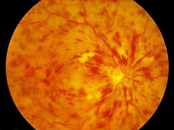
→ reduced blood flow or clot in CRV
- ATS
- HT
- Glaucoma
- leukemia + plateled dysfucon
- decr. unilat VA
- disc + retinal edema
- cotton wool + hemorrhage
- dilated tortuous retinal veins
- arterial occlusion
- diabetic retinopathy, AIDS
- (bilateral) papilledema
- neovasc. → RD + glaucoma
- vascular atrophy
- vasodil
- antiplat (aspirin)
Diabetic retinopathy is the major cause of poor vision in civilized countries The annual incidence of blindness from this illness varies between 0,02 % and 1 % .
Alterations in different biomechanical pathways as a result of diabetes have been implicated in the development of the microvasculature abnormalities that are seen in diabetic retinopathy. (Fig. 16.6)
The international standard classification of retinal detachment is:
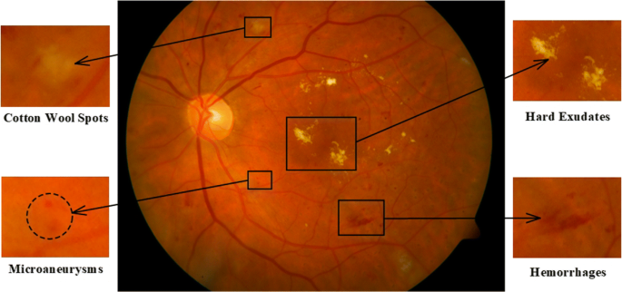
1. Background retinopathy
- dot and blot hemorrhages
- microanevrysms: outpouching of retinal capillaries which may bleed , leak, or become occluded .
- hard exudates generally most proeminant in the posterior pole. They consist in lipid components that are not easily removed by macrophages accumulate, often at the edge of the edema and are yellowish lesions .
2. Diabetic maculopathy
- macular edema
- hard exudates in complete or incomplete rings (macular star)
3. Preproliferative retinopathy
- cotton wool spots: microinfarct of the retinal nerve fibre layer causing a localized swelling of the nerve fibre layer axon.
- venous bleeding and loops: venous irregularities and engorgement .
- intraretinal microvascular abnormalities (IRMA)
- widespread capillary hiperfunction plus the findings in the first 2 stages.
4. Proliferative retinopathy:
- neovascularization within 1 disc diameter of or involving the optic disk, retina or iris. There are fragile new vessels which grow outside the retina along the posterior surface of the vitreous
- fibrous tissue along the posterior surface of the vitreous and adherent to the retina
- retinal detachment
- vitreous hemorrhage
Differential diagnosis:
- central vein occlusion
- hypertensive retinopathy
Treatment:
- Argon laser treatment in 2,3,4, stages ( panretinal photocoagulation)
- posterior vitrectomy is indicated in:
- dense vitreous hemorrhage (to clarify the vitreous )
- tractional retinal detachment (to relieve retinal traction and repair retinal detachment)
- severe retinal neovascularization and fibrous proliferation ( to eliminate the fibrous proliferation and apply further panretinal photocoagulation laser to destroy the new vessels
- backgroud - microaneurysm + dot+blot hemorrhage
- Maculopathy - edema + hard exudate
- Preproliferative - cotton wool, hemorrhages
- Proliferative - Neovascularisation → vitreous hemorrh, RD, glaucoma
- argon laser 📷
- post. vitrectomy
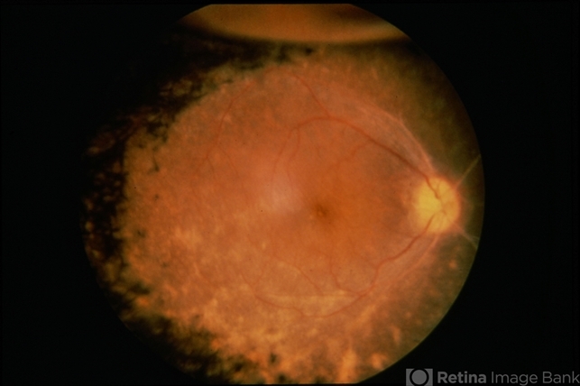
- hereditary
- rod defic.
- decr. VA at night
- loss of periph. vision
- fundus 📷 :
- periph. pigmentation (clumps + bone spicules)
- narrowing of the arterioles
- areas of depigmentation
- cataract (post. subcapsular)
- optic atrophy
Vit A (Vit E laut lecture aber suggestive contraproductive laut studien)
primary - rhegmatous (after cataract surgery, high myopia, degen. retinal lesion(retinal tear)) → check US (B-scan)
secondary - tractional, exudative, tumoral
Rest → check TR
It is defined as a separation of the pigmentary epithelium and the other embryogenetic levels
Classification:
- primary retinal detachment: - which appears on an eye with risk factors:
- aphakia
- high myopia
- degenerative retinal lesions
- secondary retinal detachment:
- exudative - in severe ocular inflammations
- tractional – in diabetic retinopathy
- tumoral – in ocular tumors
Clinical features in primary retinal detachment:
Symptoms:
- flashes of light and floaters
- decreased visual acuity in later stages
Signs:
- retinal break
- pigmented cells in the anterior vitreous
- elevation of the retinal along with a break in the retina (Fig. 16.8)
Differential diagnosis:
- secondary retinal detachment ( B-scan ocular ecography)
Treatment:
- laser
- cryotherapy
- scleral buckling surgery
- posterior vitrectomy
1-3y
- metastasis
- sec. glaucoma (neovasc.)
- cong. cataract
- prematurity retinopathy
- enucleation + excenteration 📷
- chemo
treatment → 5y
no treatment → 2-4month
- gentic + environment
- smoking
- vascular disease + HT
See Tim Root - ARMD
⇒ 📷: Algorithm Wet vs Dry ARDM
⚡ Optic nerve pathology
ant (papillitis) + post
- MS (young pat.)
- idiopathic
- infections (childhood + viral (HZ, EBV..)
- chronic infammation of sourrunding (orbital cellulitis, menigitis, sinus+teeth inflammation)
- granulomatous inflamm (tbc, syph, sarcoidosis)
- decr. VA, color vision
- pain
- compare TR
- in ant. form: 📷
- papilloedema
- peripapillary hemorrhage
- (normal in post)
- ischemic optic neuropathy
- acute papilledema (inc. ICP)
- severe HT
STEROIDS + ETIOLOGY
steroids → systemi + peribulbar
consider AB
old (>55y)
- ATS (NAION)
- giant cell arteritis (horton)
- vascular occulusion (ant or post. segment)
- sudden painless vision loss
- unilateral or bilateral
- Fundus
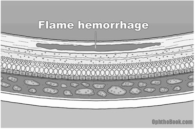
- 📷→ pale disk swelling + flame hemorrhage
- normal in post. forms
- altitudinal + central visual F defects
(vascular) optic atrophy , blindness
Giant cell → Steroids!!
vasodilators, anticoagulation, (VitB?)
- tobaco + alcohol abuse
- methyl alcohol
- etambutal (anti-tbc drug)
- DM → uremia(prob. due to RenalF → toxic subst)
- painless + bilateral vision loss (also color v.)
- Fundus
- Visual field defect
→ swelling of disc if ant. (or not if more post)
→ bilateral cecocentral or central defect

- treatment of cause (i.e. ethanol for methyl intox)
- evtl. VitB12 in chronic alcohol abuse
swelling optic nerve due to incr. ICP
BILATERAL
→ brain space occupying processes:
e.g. tumor, hematoma, hemorrhage, stenosis in CSF, abcess, inflammation (meningitis, encephalitis)
UNILATERAL
- central vein thrombosis
- orbital cellulitis
- CNS symptoms:
- normal VA (abnormal in severe + prolonged), but blurred
- Fundus → Optic disk swelling + proeminence, flame hemorrhage
→ altered mental state, headache, vommiting + nausea, vertigo
CT (or MRI)
tabes dorsalis (4th stage syphillis = neurosyphilis)
- optic neuritis
- ischemic optic neuropathy, retinal art. occulusion
- toxic optic neuropathy
- papilledema
- trauma
- tumor (pituitary adenoma)
- Glaucoma
- retinitis pigmentosa
- decr. VA
- pale disc 📷
you cant 🤡
🕳️ Pupil pathology
- leucocoria → cateract + retinoblastom a
- reddish→ vitreos hemorrhage
excetric in penetrating occular trauma
- >4mm
- spastic → sympthomimetics (adrenalin, cocaine); mediastainal tumor Pourfour de Petit sdr.
- paralytic → Paralayisis of sphincter (atropin, CN3 palsy, CNS infection, closed angle glaucoma)
- <2mm
- spastic (due to parasympthomietics or post synecchia in uveitis)
- paralytical (Horner synd → Tim Roots)
marcus gunn pupil
📷: damage visual pathyway (anywhere ganglion cell layer → lat. geniculate body)
Causes:
→ RD
→ vascular retinal artery obstruction
→ advanced glaucoma
→ optic n. pathology
- no pupillary reaction to light
- but pupillary constriction due to accomodation (near)
Causes:
- Perinaud syndrome 📷
- midbrain pathology
- compression pupillary light reflex fiber
- →large+eccentric pupil
- absent upgaze, upper lid retraction, converge, nystagmus, retraction
- Holmes Adie pupil (Adies tonic pupil) 📷
- viral infection (to ciliary ganglion)
- initially mydriatic → myosed
- near accomodation constriction delayed
- absent tendon reflex
- Miotic dystrophy
- Diabetes pupil neuropathy → light near dissociated
- Argyll Robertson pupil 📷
- dmg fibers pretectal nucleus to edinger westphal nucleus
- due to syphilis, NS tumor, chronic alcoholism, MS
- Pupills miotic, irregular, ptosis
🔪 Ocular Trauma
Watch TR → “Common Eye Trauma”
- Ecchymosis
- Excoriations
- Lacerations
- Profound or superficial wounds
- contusion (cranio-facial trauma)
- fractures wall
- optic canal (→ optic nerve lesion)
- orbital hematoma
- orbital emphysema
zones of minimal resistance → equator + limbus
erosion + abrasion
profound keratitis (into stroma) + rupture
hyphema
- subluxation of lens 📷
- luxation in AC or vitreous chamber
- concussion cataract
- chorid rupture + hemorrh
- Retinal detach, tears, edema, hemorr
- vitreous hemorrh
- eyelid → surgery
- optic nerve, muscles, vessel, foreign bodies
- conj. wounds
lens / iris prolapse → pupil distorted in shape
- ocular hypotony
- incarceration uvea
- foreign bodies
- exogenous septical uveitis
- Panuveitis
- Retain of foreign body (metal-metal injury)
surgery
(evtl. extraction of intraocc foreign body)
physical (flame, electricity)
chemical
bases
Acids = tissue COAGULATION
Bases = tissue LIQUEFACTION
- Acids are less harmful than bases due to their ability to precipitate denatured proteins, limiting tissue damage.
- Bases continue to cause damage and require continuous irrigation and pH monitoring until normalization.
- Acids have a limited impact on tissue damage, while bases have a persistent and continuous effect.
- Continuous irrigation and pH monitoring are necessary when dealing with bases to prevent further damage.
- Precipitation of denatured proteins by acids helps mitigate tissue damage.
- Bases have a relentless and continuous corrosive effect that necessitates ongoing intervention and pH monitoring.
- Grade
- Hyperemia eyelid + conj.
- Corneal erosion
- Grade
- Phlyctenules eyelid
- conjunctival erosions + edema
- cornea: abrasion, ulceration, cloudy (if stroma)
- exogenous toxic uveitis (if penetration)
- Grade
- eyelid - necrotic
- conjunctiva - pale/necrosis
- Sclera - necrotic
- exogenous uveits (as seen in G2)
- ectropion
- symblepharon
- vascularized corneal leucoma
→ heals without complications
→ evolution:
- irrigation irrigation irrigation! → wash out du schwein
- lubricant ointments
- mydriatic drops / atropine