


Thoracic Surgery
- Thoracic Surgery
- 💨 Pneumothorax
- 💥 Thoracic trauma 🔒
- 🧫 Pleural empyema 🔒
- 🌊 Malignant pleural effusion 🔒
- 🦀 Lung cancer 🔒
TRY this topic 🏎️
↓
‣
💨 Pneumothorax
‣
‣
👉🏽
Air accumulation in pleural cavity: 📷
‣
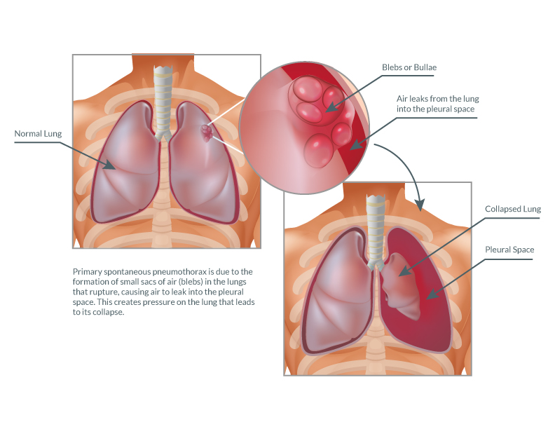
- Primary spontaneous pneumothroax (PSP)
- spontaneous
- Ø primary pathology
- <45y
- Contralateral lung normal on Xray
- Secondary spontaneous pneumothorax (SSP)
- pre-existing pathology
- pulmonary symptom preceeding PT
- >45y + smoker
- contraleteral lung with Xray abnormalities
- Iatrogenic → THoracocentesis, transbronchial biopsy, CVC, Barotrauma
- Post-traumatic
- Catamenial
‣

- PT during pregnancy
‣
- Pulmonary blebs are small subpleural thin-walled air-containing spaces, not larger than 1 or 2 cm in diameter (with the precise limit varying by source).
- Their walls are less than 1 mm thick.
- If they rupture, they allow air to escape into the pleural space resulting in a spontaneous pneumothorax.
- Blebs are a very common finding in otherwise normal individuals. They are often found in young patients. They are more common in thin patients and in cigarette smokers 1
‣
SSP → ↑mortality, more difficult treatment, longer hospitalization
‣
- COPD/emphysema
- lung cancer
- Pneumonia
- lung fibrosis
⇒ but every lung disease can cause SSP
‣
‣
👉🏽
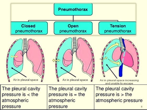
- Closed 📷
- #1
- small lesion visceral pleura
- mostly benign evolution
- Open ("sucking chest wound) 📷
- open penetrating trauma
- severe respir. insuff.
- Tension pneu 📷
- Freq. in SSP, posttraumatic or barotrauma (i.e. scuba diver)
- Severe cardio-circulatory failure (mediastinal shift)
‣
- Tension pneumothorax:
- Disruption of visceral pleura, parietal pleura, or tracheobronchial tree
- One-way valve mechanism, allowing air to enter the pleural space on inspiration but not exit on expiration
- Progressive accumulation of air in the pleural space, leading to increasing positive pressure within the chest
- Collapse of the ipsilateral lung and compression of the contralateral lung, trachea, heart, and superior vena cava
- Angulation of the inferior vena cava
- Impaired respiratory function, reduced venous return to the heart, and decreased cardiac output
- Hypoxia and hemodynamic instability
‣
T
‣
T
‣
‣
- smoking
- age
- sex
- constitutinal size (skinny+ tall)
‣
- Pleuritic pain → sudden onset
- Dyspnea (progressive)
- Cough (non-productive)
- Physical exam:
- Signs of unilateral air accumulation
- ↓/absent breath sounds
- Percussion: Tympanism/Hyper-resonance
- Tracheal deviation
- Subcutaneous emphysema
- Tachycardia+tachypena + hypotension (obstruct. shock), distended neck veins ⇒ tension P.
‣
- Xray #1 → pleural line is moved more medially and lateral part shows Øvascular signs 📷
- US → intens. pleural reflex + recurr. ecchos (A-line); loss of Sea shore sign → bar code sign; 📷
- CT → Underlying cause of SSP, esp in TRAUMA❗, DDx with emphysem bulla or ruptured pulmonary cyst
if tension p → mediastinal/tracheal deviation
‣
‣
Acute:
- reexpansion of the lung
- prevention of recurrence
Observation and Aspiration
‣
‣
- classic treatment fails
- P. recurrence
- persistent P.
- prolonged air leaks (> 5 days)
- tension P.
- bilateral P.
- professions at risk (pilot, diver)
- need for pulmonary biopsy
‣
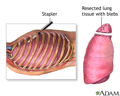
atypical resection of affected lung and partial parietal pleurectomy (VATS or thoracotomy(open) 📷
‣
severity of symptoms
‣
💥 Thoracic trauma 🔒
- Basics
- Airway obstruction
- Tension pneumothorax
- Open pneumothorax
- Massive hemothorax
- Flail chest and lung contusion
- Cardiac tamponande
‣
🧫 Pleural empyema 🔒
‣
🌊 Malignant pleural effusion 🔒
‣