


Radiology II
🫁 Lung 🔒
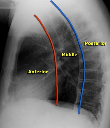
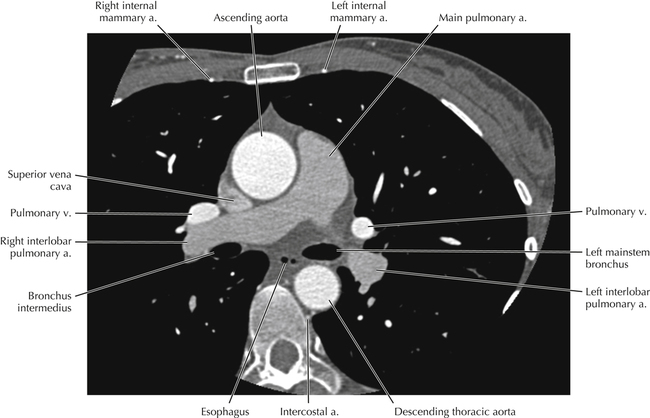
- Anatomy + Terms
- Analysis of Xray
- Pulmonary syndromes
- Pneumonia (Inflammatory non-suppurative pneumopathy)
- Bronchopulmonary tumor
🫀 Mediastinum
- SUP mediastinum ⇒ Sup. vena cava syndrome
- MIDDLE mediast ⇒ bronchi + recurr. laryngeal n. compression
- INF mediast. ⇒ Inf. vena cava syndrome
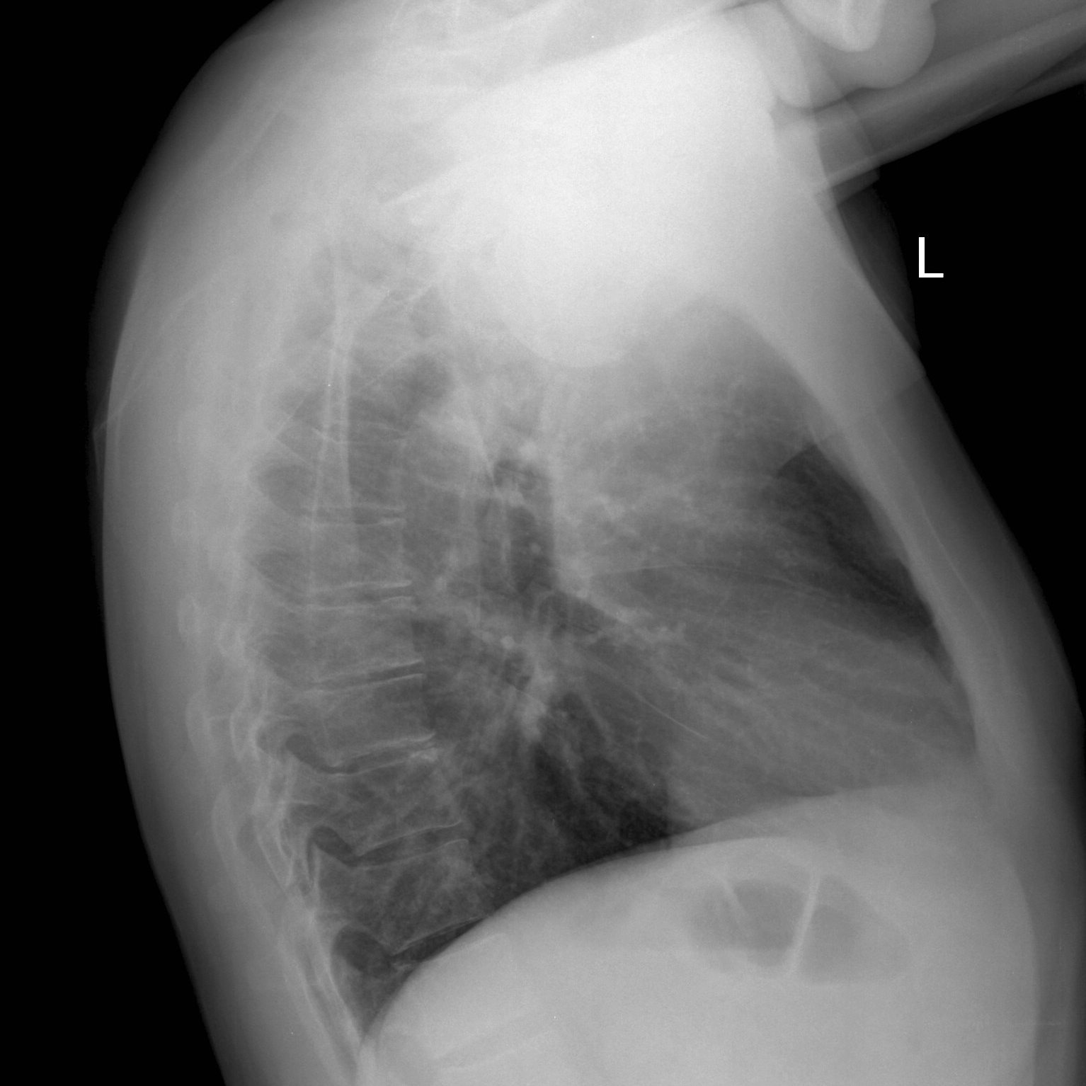
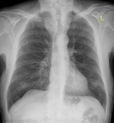
- Xray (PA, LL) → identification
- CT +con→ characterization, extension, LN, (bone involvement)
- MRI +con → Spinal / nervous system


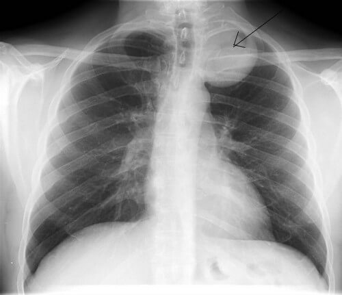
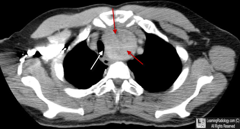
- Enlarged mass in ant. mediastinum (enhancing) [red arrows]
- Mass effect → trachea (Dislocation/compression) [white arrow]
- retrosternal goiter vs. tumor?
- homogenous → retrosternal goiter
- inhomogenous cervical LN? invasive? → tumor
scinti 📷
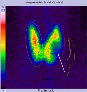
anterior middle 📷 (anterior to asc. aorta on AP view)
- Hyperplasia 📷
- Cyst 📷
- Thymoma 📷
- Carcinoma 📷
- Teratoma 📷
→ global enlarged → normal shape/outline
→ fluid density → wall calcification
→ benign features, → homogenous contrast uptake → +/- calcifications

→ inhomogenous → WELL DEFINED OR INVASIVE/mets → inhomogenous contrast uptake

→ fat + calcifications → cystic (uni or poly)
Middle mediastinum 📷
- Infectious
- Malignancy
- Primary: Lymphoma (HL+NHL)
- Secondary LN mets
- Granulomatous
- Sarcoidosis
- Silicosis (egg shell)
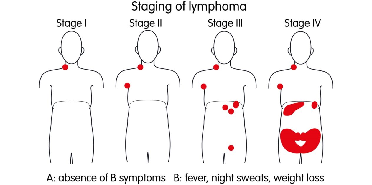
Stage | LN group | extralymphatic region |
1 | 1 ———> OR: | 1 |
2 | 2 ———>OR: 1+
(same side diaphragm) | 1
|
3 | 2
(both sides diaphragm) | +/- (spleen) |
4 | +/- | multiple organs |
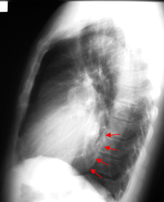
Posterior mediastinum (paravertebral) 📷
- Periph nerves → Neurofibroma + Schwannoma (Neurinoma)
- Sympathetic → Neuroblastoma
- opacity (paravertebral)
- Øsilhouette sign (behind heart)
- round/oval
Pathology of other structures in post. mediastinum
→ esophagus, thoracic aorta, spine
🫀 Heart
Soft tissue evaluation!
- MYOCARD
- MI
- hypertrophy (+wall function)
- tumor invasion (also pericardal tumor)
- SEPTUM
- defects? 📷
- Other imaging exams: echo, xray, scinti, angio, catheter
→ see below
LEFT → Backwards (retrocardial)
Right → Forwards (retrosternal)
⇒ touches >1/3 of retrosternal clearspace (inner ant. chest wall)

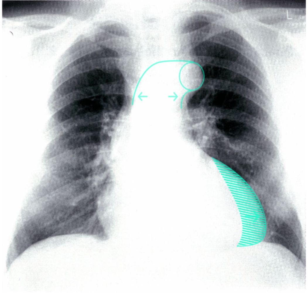
- Aortic knob: >35mm distance to lateral. air border of trachea 📷 📷
- ↑Pressure/Flow
- Aortical wall abnormalities
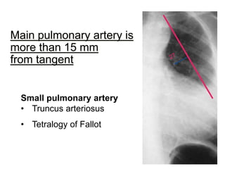
- Main Pulmonary artery (might not be seen)
→ Normal: 0-15mm medial from tangent line (aortic knob→LV) 📷 📷
→ If extends beyond (convexity?) ⇒ Pulmonary artery HT (↑Pressure/flow)
Venous → pulmonary capillary p >(10-)12
Arteral → PAP systolic >25 OR PAP median >15
- Oligemia ⇒ ↓blood into lungs (obstrucion (PA stenosis), ↓V (hypovolemia +R-L shunt)
- Hyperemia
- Venous PHT
- Arterial PHT
- venous PHT (LV congestion)
- Obstruction
- embolism
- Primary PHT
⇒↑blood into lung (PA regurg, ↑V( hypervolemia, L-R shunt)
⇒ LV congestion (mitral stenosis, LV failure)
Lung | Hili | Vessels | Felix rim (Fleischer signs) | |
Oligemia 📷 | Hyperlucent periph. (Westernmark) | small + frail | small (+ not filled) | enlarged PA 📷 |
Hyperemia 📷 | Hyperlucent (relative) | Arterial hili | dilated + redistribution (cephalization) | Ø |
mild venous PHT (12-18mmhg) 📷 | Venous hilum | visible pattern + redistribution
(cephalization) 📷 | Ø | |
moderate venous PHT
(18-25) 📷 | 1. interstital edema
→ Kerley A+ B, blurred hilum (cuffing) , vascular opacities
2. pleural effusion (free/loculated)
📷 | “ | “ | |
severe venous PHT (>25) 📷 | (acute) alveolar edema
→ air-space filling syndrome
→ central Bat/butterfly wing | “ | “ | |
arterial PHT 📷 | Hyperlucent
(periph collapse) = Westermarck sign) |
Large + „Amputated” = embolus
(Goodwin/Knuckle sign)
| ↓periph. vessels
Pulmonary artery >15mm from tangent | enlarged PA (ri. desc. pulmonary artery) 📷 |
Pulmonary Arterial Congestion | Pulmonary Venous Congestion |
Active congestion | Passive congestion |
Pulmonary arterial hypertension | Pulmonary venous hypertension |
Constricted arterial vessels | Prominence and thickening of upper lobe vessels |
Dilated hilar trunks | Prominence of lower lobed vessels |
Pulmonary vasculature on CXR findings | Hazy hilar vessels present |
Seen in ASD, VSD, PDA | Seen in left sided obstruction such as mitral or aortic valve defects regurgitation and stenosis |
Roentgen features of Kerley A, B, C lines | |
Kerley B Lines= pulmonary venous pressure is at 17- 20 mmHg | |
Pulmonary edema= > 25 mmHg |
- apex lifted boot shaped (”coeur en sabot”)
- prominent pulmonary artery (aPHT)
- retrosternal space filling (LL)
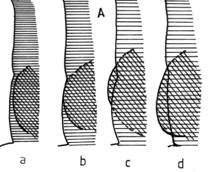
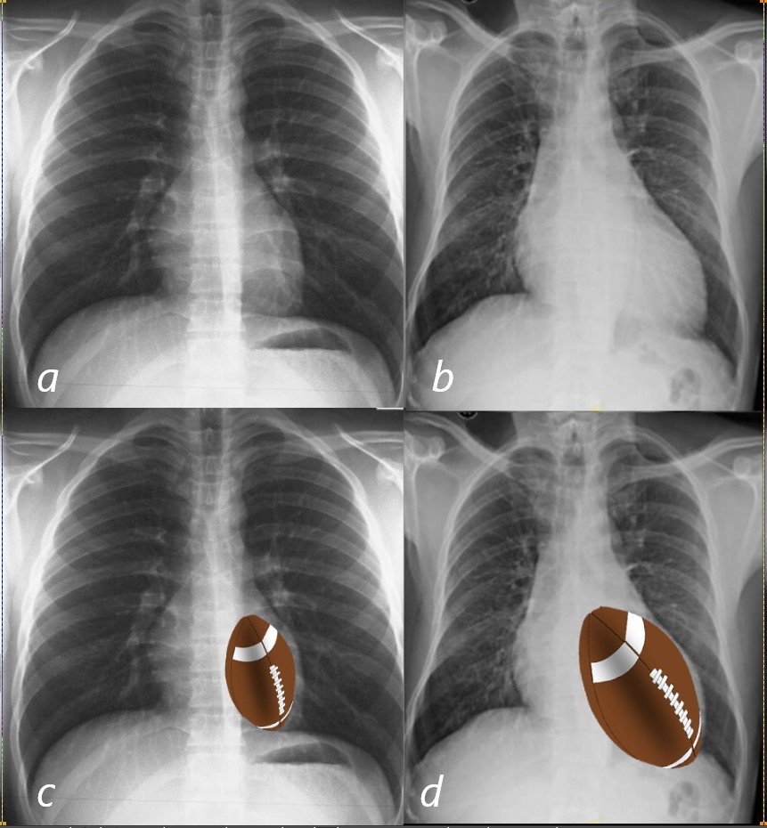
- LA enlargment
- covex/straight appendix
- double-density sign
- elevation left main bronchus + ↑carina angle
- Venous pulmonary hypertension
- upper zone venous enlaregment
- pulmonary edema
- Progresses: arterial pulmonary HT, RV hypetrophy, trikuspid regurg
- LA enlargement (see above)
- Venous pulmonary hypertension
- upper zone vein enlargemen
- acute:edema
- LV enlargement
- Enlarged LV
- prominent aortic knob (if aortic root disease)
- →concave border (tangent)
- LV enlargement (hypertrophy → later dilation)
- post-stenotic dilation
- later: pulmonary venous cong
- global enlargement (w/ clear margins)
- “lying on diaphragm” - shape change
- ↓pulsations + ↓mobility
- +/- signs of HF (clinic + radio)
- congestion (vPHT→ aPHT)
🧣 Head & Neck 🔒
- 👁️ Eye
- 🪗 Larynx
- 🦋 Thyroid
- 💀 Sinuses
🌶️ Blood vessels 🔒
- Arterial occlusion
- Aortic aneurysm + Complications
- Varicose veins
- Venous obstruction
- PE
🧠 Neuro 🔒
- Basics Neuro CT & MRI
- Stroke
- Tumors
- Trauma
- Spine
🚑 Emergencies 🔒
- Chest
- Head
- Spine