


Pulmonology
- Pulmonology
- 🏮 Bronchiectasis 🔒
- 🍏 Lung abscess
- 🪱 Hydatid cyst 🔒
- ⚰️ Sarcoidosis 🔒
- 🪘 Tuberculosis 🔒
- 😴 Sleep Apnea 🔒
‣
🏮 Bronchiectasis 🔒
TRY this topic 🏎️
↓
‣
🍏 Lung abscess
‣
‣
inflammatory circumscribed focus (nidus) which evolves towards necrosis and excavation→ bronchorrhea
‣
- primary→ pneumonia, bronchoaspiration, PE
- secondary→ bronchial obstruction
‣
- Aspiration of oropharyngeal content→ states of unconciousness, deglutition disorders, Obstruction, Ileus, Vomiting, ENT/Dental Interventions
- Hematogenous dissemination
- Pre-existing lung diseases→ischemia,necrosis
- Immune deficiencies
- Infected thorax wounds
‣
- #1 by direct inhalation - bronchial
- check for obstruction!
- parenchymal
- hematological dissemination (vasculary)
- point of entry→ ENT/ Dental/ CUtaneous
‣
‣
- non-specific: fever, asthenia, weight loss
- cough
- purulent
- fecal-smelling (anerobe)
‣
topographic stability of auscultatory signs during daily auscultation
‣
- Building-Up phase/ Closed fester→ Pneumonia signs
- Vomica phase→ exhibition of purulent, fetide and abundant sputum, fever decreases
- Open fester phase→ alteration of general condition between periods of fever and abundant sputum with no fever/small amounts of sputum
‣
Findings?:
‣
- Cultures
- Sputum examination→ collect before starting AB Tx ‼️
- Biohumoral→ Leukocystosis, Procalcitonin, Glycemia
‣
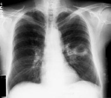
⇒ 📷
- hydroaeric levels.
- soft wall
- irregularity in hydroaeric levels in various examinations
- drainage bronchus (more visible on CT)
‣
⇒ 📷
‣
‣
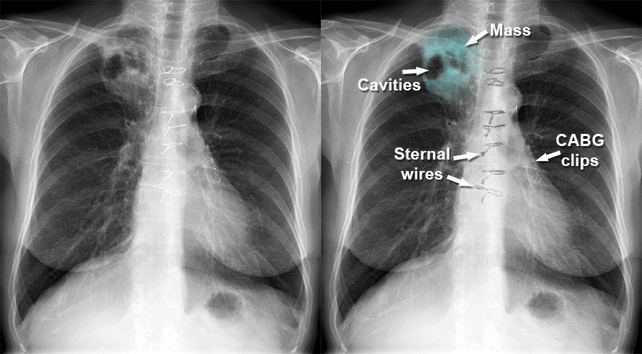
in any lung or pleural abscess→ to identify bronchopulmonary tumor
‣
- excavations of existing:
- no history of aspiration
- no fever and sputum
- no answer to ABs
- CXR
- cavity progresses towards center→ necrosis
- irregular walls
- Cytologic examination of sputum
- Fibrobronchoscopy + biopsy→ if non-conclusive→ Thoracotomy
- infectious:
- Sputum (aspergillus +)
- CT or xray ⇒ 📷
- meniscal (”c-shaped) picture
- pre-exisiting cavities
‣
‣
‣
‣
⇒ 📷
‣
→occupational exposure
‣
‣
upper lobes
‣
caverna. 📷
‣
microscopy + culture (BK+)
‣
‣
pre-existing cavities
→ evacuated hydatid cyst, emphysema air pockets
‣
‣
→ no inflammation signs around
‣
→ suspect in animal breeders (aka farmer fut)
→ ANTIBODIES IN SERUM
‣
‣
- gas bubble (stomach) + fluid levels
- double heart border
‣
endoscopy or barium swallow
‣
‣
- asphyxating of vomica
- systemic spread
- septicemia→ brain, renal abscesses, DIC
- local spread
- contralateral pneumonia
- Hemoptysis
- Pleural empyema
‣
- becomes chronic
- Bronchiectasis
‣
‣
- Infection Tx→ rapid, early, long-term
- Areal/ Causative treatment
‣
after acute phase is over
‣
- chronic lung abscess, non-responging to AB Tx>3 months
- irreversible obstruction
- abscess >6cm
- Hemoptysis
- Empyema
‣
- bronchiectassis
- retractile plachypleuritis → restrictive resp. failure
‣
🪱 Hydatid cyst 🔒
‣
⚰️ Sarcoidosis 🔒
‣
🪘 Tuberculosis 🔒
‣