


Medical Imaging
- Medical Imaging
- 🏴☠️ Conventional GI Radiology
- 🔊 Ultrasound in digestive pathology 🔒
- ⚛️ CT in digestive pathology 🔒
- 🧲 MRI in digestive pathology 🔒
- ☢️ Nuclear medicine 🔒
TRY this topic 🏎️
↓
‣
🏴☠️ Conventional GI Radiology
‣
‣
- assessment of movements of GI-T
- post-op assessment Morphology+dynamic
- chronic GI-T diseases
‣
- !perforation
- !occlusion
- recent GI-hemorr, acute digestive D, pregnancy
‣
‣
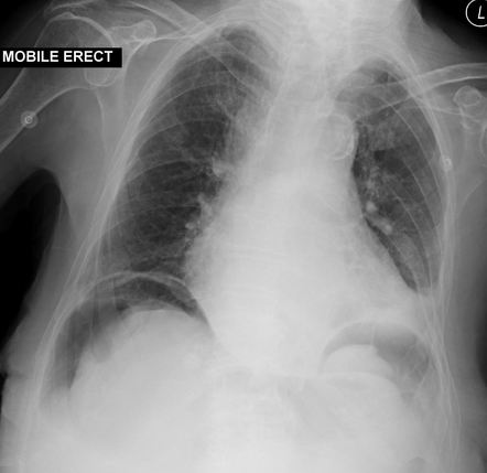
- Pneumoperitoneum
- Before contrast! → performation or obstruction??
- suspected contraindications for contrast xray: Bowel perforation, Bowel occlusion → diff. mechanical + paralytic → where is the mechanical obstruction?
- quick diagnosis of perforation or obstruction
- othercomplication
‣
‣
air → expands subdiaphragmetic 📷
‣
‣
cholecystitis, apendicitis, Ulcer, pancreatitis, diverticulitis
‣
‣
‣
‣
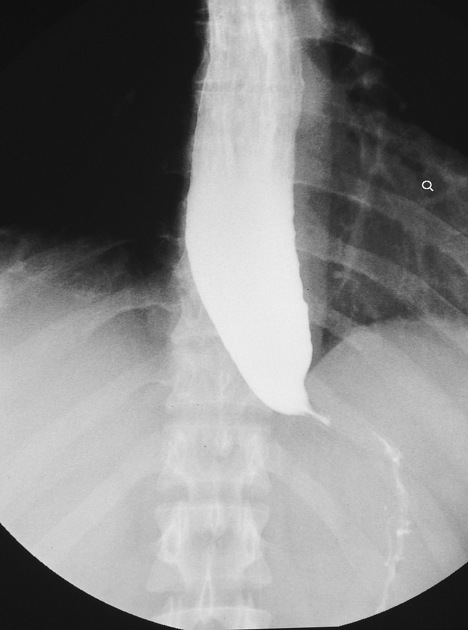
failure to relax of LES, hypertonic at rest
no primary persistaltic waves in lower 2/3
‣
👉🏽
- bird beak appearach
- no gas bubble in stomach
- dilated esophagus (megaesophagus)
- lengthenic of the esophagus
‣
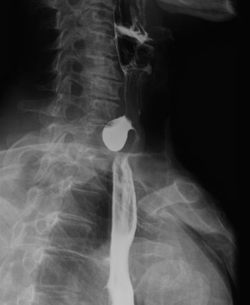
Features | Benign Stenosis 📷 | Malignant Stenosis 📷 |
Relation with the long axis | Axial, symmetrical | Usually excentric |
Junction with the normal esophagus | Progressive, smooth tapering | Abrupt, "overhanging margins" |
Length of stenosis | Spreads in length | Usually under 6 cm in length |
Contour of stenosis | Esophagitis = irregular initially; later = regular | Irregular, can contain ulcerations and lacunar images |
Localization | Above a physiological narrowing | Anywhere |
Suprajacent dilation | Always present, depends on duration | Light or Moderate |
Number of stenoses | Unique, rarely multiple | Unique |
‣
‣
‣
Criterion | Benign Gastric Ulcer 📷 | Malignant Gastric Ulcer 📷 |
Localization | Lesser curvature (vertical) | Antral region |
Size | Under 1 cm | Over 2 cm |
Shape | Round/oval | Irregular |
Contour | Smooth | Irregular |
Convergence of Folds | The folds converge till the border of the ulcer (benign relief) | The folds end abruptly at some distance from the ulcer (malignant relief) |
Structure | Homogeneous | Inhomogeneous |
Relation with the Gastric Border | Passes the gastric border | Doesn’t pass the gastric border (embedded ulcer) |
Depth | More deep than wide | More wide than deep |
Carman Ulcer | Absent | Characteristic for ulcerated cancer |
Hampton Line | Present 📷 (thin, sharp, lucent line that traverses the orifice of the ulcer) | Absent |
‣
‣
ulcerated, polypoid, infiltrative (+fungating)
infiltrative = linits plastica (=”leather bottle”, “scirrhous CA”) 📷
👉🏽
‣
‣
Characteristic | Malignant Stenosis | Benign Stenosis |
Relation with the Stomach Axis | Axial | Eccentric, towards the lesser curvature |
Length | Long | Short |
Contour | Irregular, rigid walls | Smooth, flexible walls |
‣
🔊 Ultrasound in digestive pathology 🔒
‣
- Useful artifacts
- Doppler
- Contrast agents enhanced US
- Types of US examinations
‣
- Liver Physiology
‣
- Chronic hepatitis
- Liver steatosis (fatty infiltration)
- Liver cirrhosis
‣
- Hepatic Abcess
- Hepatic congenital cysts / Biliary cyst
- Hydatid hepatic cyst
- Hepatic hemangioma (benign)
- Hepatic metastasis
- Hepatic Malignancies (lab)
- Polycystic disease (lab)
‣
- Anatomy
- Billiary Lithiasis
- Acute Cholecystitis
- Choledocal lithiasis
- Obstructice Jaundice
‣
- General US of the pancreas
- Acute Pancreatits
- Chronic pancreatits
- Pancreatic Pseudocyst
- Benign pancreatic tumor
- Malignant pancreatic tumor
‣
- Normal spleen on US?
- Splenomegaly
- Splenic hematoma
- Splenic tumor
‣
⚛️ CT in digestive pathology 🔒
- Liver pathology
- Gall bladder pathology
- Pancreas pathology
- Digestive tract pathology
- Spleen
‣
🧲 MRI in digestive pathology 🔒
- Generalities
- Hepatobiliary system with contrast
- Pancreas
- Digestive with contrast
‣
☢️ Nuclear medicine 🔒
- Basics
- Bone scintigraphy
- Renal scintigraphy